What is Sports Physiotherapy?
If you have heard of athletes using sports physiotherapy to treat or prevent injury, you might be wondering what it is. Physiotherapists are skilled at:
- Evaluating and diagnosing the injury
- Creating a personalized treatment plan
- Teaching patients methods of pain management
- Implementing treatment plans
You might have heard of physiotherapy being used to treat and rehabilitate when it comes to car accidents or medical conditions, but what does it mean in terms of sports? Ortho Neuro Physiotherapy Clinic, Vasundhara, Ghaziabad
Treatment of Athletes
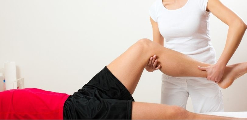
Differs from standard physiotherapy as the treatment is focused on the evaluation, prevention, management, and rehabilitation of injuries that are a direct result of exercise and sports. It can be used for athletes of all ages, abilities, and sports. Physiotherapists will often provide advice for safe practices in sports, positive lifestyle choices, as well as therapeutic treatment. Physiotherapists will help athletes improve performance as well as recover from injury, and guide them to better sports practices.
Goals of Treatment
One of the biggest differences between sports physio and other physio is the goals of the treatment. Athletes often get started with physiotherapists due to an injury. With the guidance of a physiotherapist, athletes will be treated and rehabilitated in order to not only help heal the injury, but also to prevent future injuries. The treatment is meant to help athletes return to their sport as soon as possible.
Your physiotherapist will work to protect the injured tissues in order to promote healing while helping you to control inflammation. Together, you will be able to improve your overall flexibility, strength, and muscle balance in order to safely return you to your sport and prevent you from reinjuring the area in the future.
The Process
The process of rehabilitation has several steps to get you or other athletes back to performance level. They go as follows:
To begin with, you will meet with your qualified sports physiotherapist and review your injury or concern. Through the use of several tools and evaluations, your physiotherapist will confirm the injury and help build a plan to return you back to functionality.
Once you have a plan in place, you will then begin treatment. Most often treatment is comprised of several appointments a week for several weeks. The treatment is individualized to the patient’s needs and can include several different methods, including:
- Strength training
- Ultrasound
- Stretching
- Massage
- Taping
- Body work
- Patient education
- Exercise prescription
When your injury has been properly addressed and treated, then your physiotherapist can focus on performance enhancement. The goal of this treatment is not simply to make you a better athlete, but it is to focus on the weaker or imbalanced areas on your body so that you can build up the weaker areas. This way, you will be able to prevent injuries in the future by honing in on where they are most likely to happen. Your treatment plan will be individually designed to focus on your sport, position, and any recommendations that you might need to follow to prevent future concerns.
Why See a Sports Therapist?
While you might think that all physiotherapists do the same types of treatment, a sports physiotherapist knows the details of sports and what is required to treat athletes specifically. Sports therapists know how to focus on the musculoskeletal rehabilitation that athletes need. They know that athletes want to return to their sport as quickly as possible and know what it takes to make that possible. If you are an athlete, always make sure that you see a sports physiotherapist for any treatment.
Sports Physiotherapy Common Conditio
Common Shoulder Injuries
In 2006, approximately 7.5 million people went to the doctor’s office for a shoulder problem, including shoulder and upper arm sprains and strains. More than 4.1 million of these visits were for rotator cuff problems.
Shoulder injuries are frequently caused by athletic activities that involve excessive, repetitive, overhead motion, such as swimming, tennis, pitching, and weightlifting. Injuries can also occur during everyday activities such washing walls, hanging curtains, and gardening.
Warning Signs of a Shoulder Injury
If you are experiencing pain in your shoulder, ask yourself these questions:
- • Is your shoulder stiff? Can you rotate your arm in all the normal positions?
- • Does it feel like your shoulder could pop out or slide out of the socket?
- • Do you lack the strength in your shoulder to carry out your daily activities?
If you answered “yes” to any one of these questions, you should consult an orthopedic surgeon for help in determining the severity of the problem.
Common Shoulder Injuries
Most problems in the shoulder involve the muscles, ligaments, and tendons, rather than the bones. Athletes are especially susceptible to shoulder problems. In athletes, shoulder problems can develop slowly through repetitive, intensive training routines.
Instability
Sometimes, one of the shoulder joints moves or is forced out of its normal position. This condition is called instability, and can result in a dislocation of one of the joints in the shoulder. Individuals suffering from an instability problem will experience pain when raising their arm. They also may feel as if their shoulder is slipping out of place.
Impingement
Impingement is caused by excessive rubbing of the shoulder muscles against the top part of the shoulder blade, called the acromion.
Impingement problems can occur during activities that require excessive overhead arm motion. Medical care should be sought immediately for inflammation in the shoulder because it could eventually lead to a more serious injury
Rotator Cuff Injuries
The rotator cuff is one of the most important components of the shoulder. It is comprised of a group of muscles and tendons that hold the bones of the shoulder joint together. The rotator cuff muscles provide individuals with the ability to lift their arm and reach overhead. When the rotator cuff is injured, people sometimes do not recover the full shoulder function needed to properly participate in an athletic activity.
Treatment of Shoulder Injuries
Researchers have discovered that managing your shoulder injury with physiotherapy is usually successful. Normally, you have two options: non-operative or a surgical approach. Your condition will dictate which option is best for you at this time. Non-operative care is also referred to as conservative rehabilitation.
If shoulder surgery is required, then your physiotherapist may undertake:
pre-operative rehabilitation :to either try a non-operative treatment approach or to condition and prepare your body for a surgical procedure.
post-operative physiotherapy :to regain your range of movement, strength, speed and function both to normal.
Physio Works physiotherapists have a special interest and an excellent working relationship with Brisbane’s leading shoulder surgeons to provide you with both conservative and operative rehabilitation options to ensure that you will attain the best outcome for your shoulder injury.
A common sound heard and felt within the elbow of a professional baseball pitcher. The cause is a torn ulnar collateral ligament. This elbow injury is commonly reported throughout the year during the baseball season. What goes less noticed, however, are the day-to-day aches and pains that both professional and amateur athletes go through – sprains, strains, tendonitis and bursitis. All of these can affect our performance in sport, work, and daily living.
UCL Injury
The damaged ulnar collateral ligament (UCL) can be a setback for any individual. With varying degrees of injury to the ligament,
Bursitis
If you have ever hit your elbow hard enough, especially on the edge, then you may have noticed major swelling, also known as bursitis.
Tennis Elbow
Another irritating elbow issue is best known as “tennis elbow.” In the medical arena it is known as lateral epicondylitis. This can happen to anyone from the grappler to the powerlifter and results from overuse of the extensor muscles in the forearm. The onset of pain is gradual and as it worsens the person is unable to perform his/her activity at the highest level. If caught early enough, it is best treated with rest, ice, compression, and elevation (RICE). For those who may wait a bit longer, the use of other therapeutic aides may be necessary. These aides can range from anti-inflammatory/pain medications to a brace. This is all dependent upon what instructions you are given from a medical professional.
Muscle Strains
Muscle strains can occur anywhere, lower leg to back to shoulder to anywhere there are muscles in the body. Around the elbow, the biceps, triceps, forearm flexors, and extensors are the most commonly injured. The worst of these can be a torn biceps. When this happens the muscle “balls” up in the upper arm. From here there are two options: let it heal as it is or have surgery to have it reattached. In most cases, surgery is the best option. This choice all depends on age, activity level, the individual involved, and the physician.
Rehabilitating the Elbow
Taking care of and rehabilitating the elbow is a rather different process. Most of the time, it’s all about strengthening the injured muscle. Well, being a joint, it needs strengthening both above and below the joint. For the sprained elbow, once inflammation is down and range of motion has been restored, strengthening can proceed. The best thing to do is strengthen the forearm by doing gripping exercises, such as scrunching up a towel, squeezing putty, or performing flexion and extension exercises using a dumbbell
The elbow, although a fairly stable looking joint, can be susceptible to many injuries. They can range from relatively minor to something major that requires surgery. As an athlete, it is good for you to have a basic idea of injuries and how to handle them.
Recovering From Elbow Injury
- 1. Always begin with RICE.
- 2. Move to range of motion exercises, regaining flexion and extension as needed.
- 3. Incorporate exercises that will help build strength and stability to the joint.
If ever in doubt about the injury, check with your physician. Activity should last a lifetime. Better to take care of something early than wait until it becomes a problem. Injuries are a part of life; how the injury is handled, determines how well a successful recovery will occur.
Tennis Elbow Injury Treatment
The first step is to rest your arm and avoid the activity that causes your symptoms for at least 2 – 3 weeks. You may also want to:
- • Put ice on the outside of your elbow 2 – 3 times a day.
- • Take nonsteroidal anti-inflammatory medications (such as ibuprofen, naproxen, or aspirin).
If your tennis elbow is due to sports activity, you may want to:
- • Ask about any changes you can make to your technique.
- • Check any sports equipment you are using to see if any changes may help. If you play tennis, changing your grip size of the racket may help.
- • Think about how often you have been playing and whether you should cut back.
If your symptoms are related to working on a computer, ask your manager about making changes to your workstation or have someone look at how you chair, desk, and computer are set up.
Wrist Pain
Wrist pain or injury is common. Fortunately, most wrist pain is non-life threatening and can usually be successfully diagnosed and treated by your physiotherapist or doctor. Wrist pain occurs from many potential sources of everyday life. These include sports injuries, work injuries or simple everyday arm use.
For optimal wrist pain relief and function return, an accurate diagnosis is essential. Wrist pain has multiple causes. these include wrist joint dysfunction or arthritis, wrist ligament injury, tendinopathies or muscle injury. Alternatively, wrist pain can be referred from your cervical spine (neck joint dysfunction or a pinched nerve). Local nerve compression can also occur with two common presentations: carpal tunnel syndrome or ulnar nerve palsy. Both of these conditions should prompt early assessment and treatment to avoid permanent nerve damage and loss of muscle power, sensation and function.
The good news is that most wrist pain and injury respond favourably to physiotherapy or other medical intervention. Please do not delay consulting your healthcare practitioner if you experience wrist pain. Some wrist conditions do require surgery, so early assessment and intervention is important.
Common Sources of Wrist Pain
Broken Wrist
Fractured Wrist
Common wrist fractures include:
- Fractured Radius (see image)
- Fractured Ulna
- Colles Fracture (# Radius + # Ulna)
- Fractured Carpals
- Scaphoid Fracture (most common)
- Lunate Fracture / Kienbock Disease
- Capitate Fracture
- Trapezium Fracture
- Trapezoid Fracture
- Triquetrum Fracture
- Hamate Fracture
- Pisiform Fracture
Each wrist fracture (broken wrist) needs specific rehabilitation based on injury type and fracture stability. Unstable fractures will almost always require surgical stabilization. Stable fractures will be treated with a protective and supportive splint and monitored for appropriate fracture healing. If they show signs of instability, then your surgeon may consider operative stabilisation.
Hand Swelling, Pain or Pins & Needles
Beware of CRPS (Chronic Regional Pain Syndrome)
Hand Swelling, Pain or Pins & Needles
It is extremely important to prevent hand and finger swelling post-fracture. Near permanent elevation (high arm sling) and regular finger and upper arm movement while protecting the fracture is the key. CRPS is a very significant and painful complication that can complicate your rehabilitation. Carpal tunnel syndrome which may present as pins and needles to your hand. Please report any increase in swelling, pins and needles, or pain to your physiotherapist or doctor ASAP.
It is extremely important to prevent hand and finger swelling post-fracture. Near permanent elevation (high arm sling) and regular finger and upper arm movement while protecting the fracture is the key. CRPS is a very significant and painful complication that can complicate your rehabilitation. Carpal tunnel syndrome which may present as pins and needles to your hand. Please report any increase in swelling, pins and needles, or pain to your physiotherapist or doctor ASAP.
Post-Fracture Exercises
Post-fracture exercises are specific to your fracture and should be performed after assessment and guidance from your healthcare professional. Based on that, it is very important to only perform the exercises prescribed by your Orthopaedic Surgeon or Physiotherapist. You should also wear your wrist split/cast at all times unless advised otherwise by your physiotherapist or surgeon.
Do your exercises on a frequent basis throughout the day. Multiple sessions of short duration are generally better than longer sessions done only once or twice.
For more specific advice please consult your physiotherapist or doctor.
Muscle Strain
What is Myalgia?
The most common source of myalgia (or muscle pain) is a muscle strain due to overstretching. A muscle strain may also be referred to as a pulled muscle or a muscle tear. A muscle strain can vary in severity from mild to severe and ultimately, a complete muscle rupture. Either way, your muscle strain will result in muscle pain, or at least muscle soreness, plus reduced muscle function including muscle weakness, stiffness or tightness.
What is a Muscle Strain?
Commonly, your muscle strain will occur at high-speed when your muscles are overloaded. The most common high-speed muscle injuries occur in your hamstrings (hamstring strain), quadriceps (thigh strain), calf (calf muscle tear), back (back muscle pain), groin strain. But, obviously, any muscle in your body is susceptible to a muscle strain or tear. You can also suffer a fatigue-related muscle strain from sustained postures. Back muscle strain, shoulder and neck muscle strains are often postural fatigue-related muscle strains. Text neck has become a common condition due to postural fatigue while overusing your phone. Another common source of myalgia can be related to excessive muscle microtrauma from an overdose of exercise. This condition is known as DOMS or delayed onset muscle soreness. Other overuse injuries such as RSI – repetitive strain injury, results from a combination of postural fatigue and overuse of the smaller upper limb muscles. Luckily, early assessment and prevention strategies have virtually eliminated RSI from offices in the western world.
What are the Characteristics of a Muscle Strain?
Muscle strains have the following symptoms:
- Muscle tightness
- Bruising
- Weakness
- Inability to fully stretch your injured muscle
Muscle Strain Treatment
Muscle strain treatment will vary depending upon an accurate diagnosis from your health professional. The severity of your muscle strain, and what function or loads your injured muscle will need to cope with, will impact the length of your healing and rehabilitation process.
Until you’ve been accurately diagnosed with a muscle strain, use the following guidelines:
- Ice and a compression bandage.
- Elevate the injured region if it is swollen.
- If it’s painful to walk you should be using crutches.
- Cease or reduce your exercise or activity level to where you feel no pain. Muscle strain can take anywhere from a few days to several weeks to rehabilitate successfully. Please seek the advice of your physiotherapist, doctor or your health care practitioner who specialises in muscle injuries eg massage therapist, to guide your treatment.
Carpal Tunnel Syndrome
What is Your Carpal Tunnel?
Your carpal tunnel protects vital structures such as the median nerve, blood vessels and tendons as they pass to and from your hand. The palm side of your wrist has a band of strong ligaments (flexor retinaculum) that attach to the carpal (wrist) bones at either side. The rear of the tunnel is a curved compilation of the wrist bones.
Potential Compression Sources
Your symptoms can originate from elsewhere along the median nerve. This source is frequently overlooked and could save you from unsuccessful surgery. Your lower cervical spine especially C6, C7, C8 and T1 should be thoroughly examined by your physiotherapist. This is version “double crush syndrome” and there is an increased likelihood of carpal tunnel syndrome in these patients. Kwon et al (2006).
Abnormal Neurodynamics
Your nerves should freely travel along their pathways between your spine and your fingers. Any interference of their slide mobility could cause symptoms eg scar tissue, tight muscles. Your physiotherapist can assess your neurodynamics for abnormalities. Tal-Akabi & Rushton (2000).
Hormonal Factors
Hormone imbalances can cause swelling of the hands and feet, as evidenced by the condition’s prevalence in middle-aged or pregnant women.
Gripping, Repetition and Microvibration
Occupations associated with repetitive wrist flexion and extension activities, vibratory tools, and gripping have a high incidence of carpal tunnel syndrome.
What’re the Symptoms of Carpal Tunnel Syndrome?
Carpal Tunnel Syndrome (CTS) sufferers will usually experience the following symptoms in their hand or fingers:
- hand pain or aching
- pins and needles
- numbness esp at night of with wrist flexing
- burning
- weakness or cramping
- perceived swelling
Hand or Wrist Arthritis
Hand and Wrist Arthritis
Hand and wrist osteoarthritis is a common source of pain as you age.
Basically, everyday wear and tear damage your hand and wrist joints. The chances of you suffering hand arthritis increase if you have overstressed your hand joints or have experienced excessive weight-bearing activities eg boxers and gymnasts.
What is a Cramp?
A cramp is defined as a spontaneous or involuntary electrical activity of a large number of skeletal muscle fibres that quickly develops into a painful, sustained contraction (muscle spasm).
What Causes a Cramp?
Although similar in presentation, the cause of both EAMC and nocturnal cramps differ.
Unfortunately, the exact cause of a cramp is not fully understood but there are a number of hypothesis for both EAMC and nocturnal cramps as outlined below.
Treatment for Cramps
The first and foremost priority of treatment for cramps is identifying the root cause of your cramps. Whether this be tight muscles, over-exercising or a cause that requires further investigation. Our physiotherapists are experienced in finding out what it is that is causing your cramps and will point you in the right direction.
Depending on what our physiotherapists find, treatment may involve:
- Lengthening of hypertonic muscles.
- Restoring joint range of motion.
- Strengthening weak muscles.
- Lifestyle modifications:
- Fluid intake.
- Reducing coffee/alcohol intake.
- Vitamin/electrolyte balancing.
- Modifying current exercise regimes.
What is an Overuse Injury?
Overuse injuries refer to injuries sustained from a repeated action (such as repetitive strain injury) as opposed to acute injuries, which occur in an instant (such as a sprained ankle).
How to Prevent an Over use Injury
We can prevent overuse syndromes. Some of the ways to prevent this injury include:
- Warm-up (including stretching) and warm-down (including stretching) before and after all exercise.
- Use proper equipment (e.g. jogging shoes for jogging, a racquet that is the right size with the proper grip size and strings strung to your level of play).
- Increase at a rate no faster than 10% increase per week (distance, speed, weight, etc).
- Practice and concentrate on correct technique.
- Condition yourself for 2-3 weeks before starting – strength and flexibility.
- Listen to your body – pain is a warning that something is wrong. Early identification and treatment will allow you to continue your activity.
- Identify and correct the cause of pain or discomfort.
- Ensure full injury rehabilitation, e.g. a sore right leg can cause an overuse injury in the left through compensation.
Lower back pain
This a common disorder involving the muscles, nerves and bones of the lower back. Symptoms can vary from a dull ache to a sharp sudden pain. There are several different causes of low back pain, many of which are discussed below.
As always our approach at Bradley Physio is hands-on wherever possible. Following a thorough verbal and physical examination your physiotherapist may decide to use manual therapy treatments including deep soft tissue massage, spinal mobilisations and manipulation (where appropriate). Other treatments include lumbar traction, electrotherapy, and acupuncture. Postural and preventative advice, as well as a home exercise programme is recommended too. Many clients find our pilates classes are a great, long term way to manage their neck pain and prevent recurrence.
NOTE If you are not making the progress we would expect and your physiotherapist thinks you will benefit from further investigations (x-rays,scans) and/or onward referral to a Neurologist or Spinal Orthpaedic Specialist the necessary arrangements can be made by our team on your behalf.
- Neck pain
- Sciatica
- Prolapsed disc
- Whiplash
- Spondylitis/Spondylosis
- Degenerative changes
- Trapped nerves
- Headaches/Migraines
NECK PAIN
- • A stiff neck is typically characterised by soreness and difficulty moving your neck, especially when trying to turn your head to the side. It may also be accompanied by a headache, neck pain, shoulder pain and arm pain.
- • Neck pain including whiplash and headaches is a common complaint experienced by our clients. Following a thorough verbal and physical examination your physiotherapist may decide to use hands-on treatments including deep soft tissue massage, spinal mobilisations and manipulation (where appropriate). Other treatments include cervical (neck) traction, electrotherapy, and acupuncture. Postural and preventative advice, as well as a home exercise programme is recommended too. Many clients find our pilates classes are a great, long term way to manage their neck pain and prevent recurrence.
- • NOTE If you are not making the progress we would expect and your physiotherapist thinks you will benefit from further investigations (x-rays,scans) and/or onward referral to a Neurologist or Spinal Orthpaedic Specialist the necessary arrangements can be made by our team on your behalf.
Sciatica
Sciatica describes pain felt along the sciatic nerve, which runs from your lower back, down through the buttock, back of the thigh, into the lower leg and foot. It is normally the result of pressure or inflammation around the spinal nerve root as it leaves the spinal column.The sciatic nerve is the longest nerve in the body. Irritation or pinching of your sciatic nerve can cause severe back and leg pain, muscle weakness, affect mobility and can cause pins and needles or numbness in the foot.
Following a thorough verbal and physical examination your physiotherapist may decide to use hands-on treatments including deep soft tissue massage, spinal mobilisations and manipulation (where appropriate). Other treatments include lumbar (low back) traction, electrotherapy, and acupuncture. Postural and preventative advice, as well as a home exercise programme is recommended too. Many clients find our pilates classes are a great, long term way to manage their neck and low back pain and prevent recurrence.
NOTE If you are not making the progress we would expect and your physiotherapist thinks you will benefit from further investigations (x-rays,scans) and/or onward referral to a Neurologist or Spinal Orthpaedic Specialist the necessary arrangements can be made by our team on your behalf.
Prolapsed disc
Also known as slipped disc, bulging disc & herniated disc A prolapsed disc is a common injury sustained to the intervertebral discs within the spinal column. These discs lie between the vertebrae (backbones). A disc prolapse can occur in your lumbar spine (lower back), thoracic spine (upper back) or your cervical spine (neck). When a prolapsed/slipped/bulging/herniated disc occurs it is typically due to accumulated microtrauma, and the pain can be sudden and unexpected. This occurs when the jelly like centre of the intervertebral disc bulges outwards towards the spinal nerves as they leave the spinal cord. The damaged disc and the pressure on the nerve can lead to pain. Symptoms are often aggravated when bending forward, sitting, coughing, sneezing or lifting. Pain can radiate away from the spine into the arms or legs. In some cases patients will experience pins & needles, muscle weakness or numbness in parts of the limbs.
Following a thorough verbal and physical examination your physiotherapist may decide to use hands-on treatments including deep soft tissue massage, spinal mobilisations and manipulation (where appropriate). Other treatments include traction, electrotherapy, and acupuncture. Postural and preventative advice, as well as a home exercise programme is recommended too. Many clients find our pilates classes are a great, long term way to manage their neck and low back pain and prevent recurrence.
NOTE If you are not making the progress we would expect and your physiotherapist thinks you will benefit from further investigations (x-rays,scans) and/or onward referral to a Neurologist or Spinal Orthpaedic Specialist the necessary arrangements can be made by our team on your behalf.
Spondylitis/Spondylosis
Spondylosis/Spondylitis is inflammation of the vertebrae in the spine and can cause neck and low back pain. It is a general term used to describe changes that can occur along any area of the spine. It is a condition in which your discs degenerate, losing their flexibility and height to cushion the spine. Usually pain begins in the lower back or neck and as the condition progresses pain may radiate into the arms or legs. It is often described as “pressure” or “burning” pain and you may also feel tingling or numbness in your leg or foot.
Many patients find that heat is extremely effective way to relieve discomfort. Following a thorough verbal and physical examination your physiotherapist may decide to use hands-on treatments including deep soft tissue massage and spinal mobilisations. Other treatments include traction, electrotherapy, and acupuncture. Postural and preventative advice, as well as a home exercise programme is recommended too. Many clients find our pilates classes are a great, long term way to manage their neck and low back pain and prevent recurrence.
NOTE If you are not making the progress we would expect and your physiotherapist thinks you will benefit from further investigations (x-rays,scans) and/or onward referral to a Neurologist or Spinal Orthpaedic Specialist the necessary arrangements can be made by our team on your behalf.
Degenerative changes
Also known as osteoarthritis of the spine, this process is age-related ‘wear and tear’ that can affect the discs and bones of the spine (vertebrae) and the surrounding ligaments. Symptoms include early morning stiffness of the neck or low back, aching pain and reduced range of movement of the affected area. Degenerative changes (wear and tear) can be caused by the breakdown of the discs that separate the bones of the spine. As you age the spine begins to show signs of wear and tear as the discs dry out and shrink. These age related changes can cause arthritis, disc herniation and in more advanced cases, spinal stenosis (narrowing of the spinal canal).
Many patients find that heat is extremely effective way to relieve discomfort. Following a thorough verbal and physical examination your physiotherapist may decide to use hands-on treatments including deep soft tissue massage and spinal mobilisations. Other treatments include traction, electrotherapy, and acupuncture. Postural and preventative advice, as well as a home exercise programme is recommended too. Many clients find our pilates classes are a great, long term way to manage their neck and low back pain and prevent recurrence.
NOTE If you are not making the progress we would expect and your physiotherapist thinks you will benefit from further investigations (x-rays,scans) and/or onward referral to a Neurologist or Spinal Orthpaedic Specialist the necessary arrangements can be made by our team on your behalf.
Trapped nerves
A trapped nerve, pinched or compressed nerve is a term used for the compression or irritation of nerve roots exiting the spinal cord and can be due to a prolapsed/slipped/bulging/herniated disc, Other causes may be from pressure from bony nodules that form as part of the aging process. Since the nerve is unable to convey information to the brain in the normal way you may notice that although your pain may originate in one area such as you neck or low back where the nerve is trapped, the pain will often radiate into other areas of your body (arms/legs). This is known as referred pain and can be felt as pain, pins and needles or numbness. This pain is often severe, sharp or a burning type sensation.
NOTE If you are not making the progress we would expect and your physiotherapist thinks you will benefit from further investigations (x-rays,scans) and/or onward referral to a Neurologist or Spinal Orthopaedic Specialist the necessary arrangements can be made by our team on your behalf.
Headaches/Migraines
Pressure on the nerves as they exit the spinal canal in the upper neck is a common cause of cervicogenic headaches & migraine. These nerves supply the top and sides of the head, the face, ears and base of the skull. This pain can be severe and debilitating.
Following a thorough verbal and physical examination your physiotherapist may decide to use hands-on treatments including deep soft tissue massage, spinal mobilisations and manipulation (where appropriate).
NOTE If you are not making the progress we would expect and your physiotherapist thinks you will benefit from further investigations (x-rays,scans) and/or onward referral to a Neurologist or Spinal Orthpaedic Specialist the necessary arrangements can be made by our team on your behalf.
Neck and Back Injuries Treatment
- • Immobilize the head, neck, and back. Place soft packing on either side of the head to prevent side-to-side motion. Clothing or towels are handy for this purpose.
- • If the victim needs to vomit, roll the head, neck, and body as a unit so the person rests on his or her side (preferably, one person should control the head of the person with the injury while another person rolls the shoulders and hips).
- • Monitor the victim for loss of movement and development of numbness.
- • Move the person only when necessary to preserve life.
- • CPR may be required.
What Causes Hip Pain?
Hip pain is common and spread across all age groups. The hip joint and its integration with your pelvis, SIJ and lumbar spine (lower back) make it a complex region to correctly analyse and assess any dysfunction.
Hip pain can also be associated with reduced balance. A thorough balance assessment may be required to predict a falls risk. Falls prevention exercises may be prescribed by your physiotherapist to address any individual deficits. They may even advise you to utilise a walking assistance device such as a walking stick, crutches or a walking frame.
hip physiotherapist. Biomechanical deficits and subtle hip weakness that may only show on a slow-motion video are just two of the potential causes of sporting hip injuries.
Groin Pain
Groin pain is one of the most common symptoms associated with hip joint pathologies such as hip osteoarthritis and hip labral injury. There are also many other causes of groin pain that need to be excluded by a health professional. More info: Groin Pain.
Only after a thorough hip assessment will your hip pain be effectively rehabilitated to relieve your current hip pain and joint dysfunction, plus prevent the return of any future hip pain.
Common Sources of Hip Pain
Hip Joint Pain
- Hip Arthritis – Osteoarthritis
- Hip Labral Tear
- Hip Pointer
- Femoroacetabular Impingement – FAI
- Perthes Disease
- Slipped Femoral Capital Epiphysis
- Stress Fracture
- Avascular Necrosis of the Femoral Head
Hip Arthritis
- Hip arthritis commonly describes the most common for of hip arthritis, which is known medically as hip osteoarthritis.
- Hip osteoarthritis is a joint disease that mostly affects your hip joint cartilage. Articular cartilage is the hard slippery surface that covers the sections of bones that move against each other in your hip joint.
- Healthy articular cartilage allows your hip joint bones to smoothly and painlessly glide over each other and also helps to absorb any shock forces not dispersed by your hip muscles.
What’s the Treatment of Hip Arthritis?
PHASE I – Pain Relief & Protection
- Managing your hip pain. Hip pain is the main reason that you seek treatment for hip arthritis.
- Regular application of ice packs is highly recommended to reduce your hip pain.
- Your physiotherapist will use an array of treatment tools to reduce your hip pain and inflammation. These may include ice, electrotherapy, acupuncture, unloading taping techniques, soft tissue massage and temporary use of a mobility aid (eg cane or crutch) to off-load the affected side.
PHASE II – Restoring Normal Hip ROM, Strength
As your hip pain and inflammation settles, your physiotherapist will turn their attention to restoring your normal hip joint range of motion, muscle length and resting tension, muscle strength and endurance, proprioception, balance and gait (walking pattern).
Hip researchers have discovered the importance of your hip muscle recruitment patterns with a normal order of deep, then intermediate and finally superficial muscle firing patterns in normal pain-free hips. Your physiotherapist will assess your muscle recruitment pattern and prescribe the best exercises for you specific to your needs.
PhysioWorks has developed a “Hip Core Stabilisation Program” to assist their patients to regain normal hip muscle control. Please ask your physio for their advice.
PHASE III – Restoring Full Hip Function
The final stage of your hip arthritis rehabilitation is aimed at returning you to your desired activities. Everyone has different demands for their hips that will determine what specific treatment goals you need to achieve. For some people, it may be simply to walk around the block. Your physiotherapist will tailor your hip rehabilitation to help you achieve your own functional goals.
What Else Can You Do For Your Hip?
Lose Weight
Lose weight. You won’t just look better, you’ll feel better, too. Why? Every extra kilogram you carry around translates to added stress to your hip joints. Excess weight can mean more hip pain, no matter which form of arthritis you have.
Ice it!
When your hip joint is hot and inflamed, applying something cold can decrease pain and swelling by constricting blood vessels and preventing fluids from leaking into surrounding tissues.
Medications
Follow your doctor’s advice. Some medications will be designed for pain relief and others to reduce inflammation. Since most hip osteoarthritis sufferers normally have other medications, it is always wise to check with your doctor before changing.
Exercise – Keep Moving
Exercise helps to lessen your hip pain, increase your hip joint range of movement, reduce fatigue and help you feel better overall.
A well-rounded workout routine for people with hip osteoarthritis includes flexibility exercises to increase your hip joint and muscle range of motion, aerobic exercises to improve your endurance and decrease fatigue, and strengthening exercises to improve your muscle endurance and power.
Your physiotherapist is an expert in the assessment and prescription of hip arthritis exercises. Please ask them what is best for you.
General exercise such as swimming, hydrotherapy, Tai Chi, yoga, pilates, balance and walking programs are excellent if pain-free.
The key is to have a regular daily exercise program. The goal is to keep moving.
Strengthen Your Bones
Ask your doctor to check your bone density. If they are concerned they’ll arrange for a test to check if you have osteoporosis (bone thinning).
Follow their advice or the advice of your dietitian on your Calcium and Vitamin D intake. You need normal levels of both plus some form of weight-bearing exercise to strengthen your bones.
Treat Your Muscles with a Massage!
A quality remedial massage may be just the relief your hip muscles need. Treat yourself to a good rub down with someone you trust. The benefits vary from person to person but may include decreased pain and muscle stiffness associated with your arthritis, increased circulation, and an improvement in your sleep and immune functions. Mentally, massage can also decrease stress and depression.
Besides all it that, massage just feels good!
For more advice, please ask your physiotherapist, massage therapist or doctor.
Hip Pointer
What is a Hip Pointer?
A hip pointer is a contusion (bruise) on the iliac crest (top of hip bone) or across the greater trochanter (most prominent aspect on the outside of hip) as a result of direct trauma. Sometimes this can be accompanied by an avulsion injury, where a small fragment of bone is torn away by the attached muscle. Bleeding and swelling are a result of the injury and cause pain with hip movements.
What Causes a Hip Pointer?
A hip pointer injury is usually the direct result of a blow to the hip (iliac crest or greater trochanter) or from a fall onto a hard surface.
How is a Hip Pointer Treated?
Initially, a hip pointer is treated using rest, ice and compression. Ice can be applied for 15-20mins every 2-3 hours in the initial 24-72 hours following injury. A hip pointer requires adequate recovery time to allow the injured structures to heal. If walking is difficult crutches may be supplied to allow for mobilisation. Return to play will be determined by pain levels, hip mobility and based on your previous level of function. It may take 1-3 weeks to heal.
While you are waiting for the contusion to heal your physiotherapist will provide you with some simple exercises to maintain hip joint range of motion and prevent stiffness. Aquatic-based exercises can be helpful in maintaining hip joint range whilst unloading the area.
What is Femoroacetabular Impingement?
Femoroacetabular Impingement (FAI) is a hip condition which describes a mechanical mismatch between the hip “ball” and the “socket”.
There are three described types of femoroacetabular impingement:
Cam Type FAI
‘Cam’ type femoroacetabular impingement describes a ‘bump’ on the surface of the femoral head (ball) which jams on the rim of the (acetabulum) socket. This typically affects young athletic men.
Femoroacetabular Impingement Treatment
An initial trial of non-operative treatment is advocated for most patients, as the pain is relatively self-limiting.
Physiotherapy can assist FAI by using a variety of techniques to:
- mobilise the hip joint that stretches any tight structures eg joint capsule or muscles
- improve soft tissue flexibility and length
- strengthen the deep, intermediate and superficial hip muscles
- progress hip muscle, proprioception, joint position sense, and functional control to dynamically control your hip
Use of painkillers and anti-inflammatories may temporarily help the pain reduce the local anti-inflammatory reaction.
Recovery from hip arthroscopy typically takes 3-4 months, while open hip debridement is typically 12 months. Hip arthroscopy has been the preferred method in recent years and has reported excellent results with 80% of patients asymptomatic by 3-4 months and up to 95% had improved symptoms by one year.
For more advice about femoroacetabular impingement, please ask your physiotherapist or doctor.
Perthes Disease
What is Perthes Disease?
Legg-Calve-Perthes Disease, or Perthes Disease for short, is a rare condition that affects children. It affects boys more than girls and usually between the ages of 4-10 years old. It affects the hip joint and involves the femoral head (the ball part at the top of the thigh bone). The blood supply to this area of the bone becomes interrupted, with insufficient blood flow the bone begins to die (avascular necrosis). This results in a flattening of the femoral head. In most cases, this only occurs on the one side.
What is a Stress Fracture
Stress Fracture
One of the most common injuries in sport is a stress fracture. Stress fractures are tiny cracks in a bone. Stress fractures are caused by the repetitive application of force through the bone that isn’t strong enough. Essentially, the bone is weaker than is required for the activity demands or exercise intensity.
The most common stress fractures occur in runners, but stress fracture can occur due to the demands of your sport. eg lumbar spine stress fractures in gymnasts and cricket bowlers. Common running stress fractures include foot (navicular, metatarsal), tibia (shin splints). Stress fractures also can arise from normal use of a bone that’s been weakened by a condition such as osteoporosis.
What is Avascular Necrosis of the Femoral Head?
Avascular Necrosis (or Osteonecrosis) is a condition affecting the upper part of the leg – specifically the femoral head. Essentially, the femoral head or ball of your hip joint receives less and less blood supply. Since bone is living tissue when this blood supply is reduced enough the bone dies. Once the bone dies, the femoral head collapses and if severe enough, the hip joint itself collapses.
Avascular Necrosis mainly affects those aged between 20-50 years old. In general, the healthier you are, the less risk you have of developing avascular necrosis. It usually comes about secondary to an underlying health issue or previous injury.
What are the Symptoms of Avascular Necrosis?
Avascular Necrosis requires time for its onset. It is relatively asymptomatic to begin, but as it progresses, the pain becomes more pronounced. Pain limited hip range of motion is the ultimate symptom both passively and actively which can refer pain down the length of the leg. Generally, the pain will arise when pressure is placed on the bone which unfortunately includes walking around!
Avascular Necrosis can often mask itself as other conditions in the early phase given its general pain presentation within the hip. Therefore, it is paramount for your health practitioner to record a detailed history of your presentation and perform a thorough examination to identify if avascular necrosis is your true cause of pain.
Avascular Necrosis Treatment
Conservative treatment with physiotherapy has proven to be relatively ineffective for avascular necrosis. Although the aim of conservative treatment is to decrease the weight bearing load through the head of the femur (usually by implementing crutches), research has shown that the condition still progresses in 1-2 years. Without definitive treatment, 70% to 80% of all avascular necrosis of the femoral head cases will progress and inevitably undergo surgery.
Irrespective of what surgery is undertaken, the primary goal is always to preserve the natural femoral head as opposed to replacing it.
There have been proposed nonoperative treatments that can be implemented before the condition progresses and the femoral head collapses as seen in the picture on the first page. They are, however, still in the early stages of research and development. The use of various electromagnetic, acoustic stimulation or pharmaceutical modalities has been trialled with varying success. One of the most well known and successful treatments, if the condition is identified early enough, is the core decompression – i.e. decrease the pressure inside the femoral head. This is achieved through drilling holes into the femoral head to create channels for new blood vessels to nourish the affected areas of the hip or the channels are filled with healthy bone from another part of the body. This has proven to be successful, especially in those with early stages avascular necrosis based on preoperative and postoperative MRI studies.
As evident, there is a multitude of different approaches that can be taken to manage avascular necrosis. Ultimately, an orthopaedic opinion is paramount to ensure the condition is treated as effectively as possible. The decision as to which treatment to select will depend on the stage the avascular necrosis in combination with the clinical evaluation of the patient.
Knee Pain
Knee pain or knee injuries are extremely common, and there are many causes. It is important to make an accurate diagnosis of the cause of your knee pain or injury so that appropriate treatment can be directed at the cause. Knee pain can arise from soft tissue injuries eg ligament sprains and muscle strains, bone conditions eg knee arthritis, Osgood Schlatters, and biomechanical dysfunction eg Patellofemoral syndrome. It may even be referred from your sciatica!
Knee pain has many causes and your knee treatment varies considerably depending on an accurate diagnosis. Treatment can involve simple knee mobilisation techniques, massage, taping, stretches or strengthening exercises all the way through to a thorough rehabilitation protocol post knee reconstruction or knee replacement.
How Do You Know If Your Knee Injury Is Serious?
While it is always best to seek the professional advice of a highly skilled practitioner who is trained in knee injuries such as your doctor or physiotherapist, here are seven signs that could indicate a severe knee injury.
- 1. Obvious deformity. You may have a fracture or dislocation.
- 2. You heard a “pop” or “snap”
- 3. You’ve experienced swelling
- 4. Greater than normal movement
- 5. Less than normal movement eg can’t straighten
- 6. You are unable to weight-bear on your leg
- 7. Your knee “gives way” or “buckles”.
If you experience any of these symptoms please seek prompt medical assessment. Please consult your physiotherapist or doctor for the most accurate diagnosis and best treatment for your knee pain.
What is the Best Exercise for Knee Pain?
It may seem a little contradictory, but researchers have identified that knees exercises may assist the relief of your knee pain. The important thing to identify is which knee exercises are likely to help you and which could be harmful.
What exercise dosage you should be doing is also important. Your exercise dosage will vary depending upon the phase of your specific diagnosis, your knee injury diagnosis, plus other individual health factors. This is exactly where your physiotherapist’s professional training including knee injury diagnosis and appropriate knee exercise prescription is important to guide you quickly back to pain-free knees.
While we’d like to say that all knee exercises are beneficial to you, there are significant individual differences between all patients who present with knee pain. For example, an older diabetic overweight patient will require a very different set of knee exercises to a young high-performance athlete, or a patient who has just had a knee operation.
Based on the significant individual differences of who presents with knee pain, it is highly recommended that you seek the professional advice of your trusted physiotherapist or healthcare practitioner who has a special interest in knee pain and injuries to guide your knee rehabilitation.
Knee Pain Common Causes
Knee Ligament Injuries
What is a Knee Ligament?
A ligament is a short band of tough fibrous connective tissue composed mainly of long, stringy collagen molecules. Ligaments connect bones to other bones in and around joints. They do not connect muscles to bones; that is the function of tendons. Ligaments limit the amount of mobility of a joint, or prevent certain movements altogether.
What Causes Knee Ligament Injuries?
You can injure a ligament through a sharp change in direction, landing wrong from a jump, or the most common a blunt force hit to the knee, such as in football tackle. The incident usually needs to happen at speed. Muscle weakness or incoordination predispose you to a ligament sprain or tear.
How Do You Know If Your Knee Injury Is Serious?
While it is always best to seek the professional advice of a highly skilled practitioner who is trained in knee injuries such as your doctor or physiotherapist, here are seven signs that could indicate a severe knee injury.
- 1. Obvious deformity. You may have a fracture or dislocation.
- 2. You heard a “pop” or “snap”
- 3. You’ve experienced swelling
- 4. Greater than normal movement
- 5. Less than normal movement eg can’t straighten
- 6. You are unable to weight-bear on your leg
- 7. Your knee “gives way” or “buckles”.
If you experience any of these symptoms please seek prompt medical assessment
Symptoms & Severity of Knee Ligament Injuries?
The severity and symptoms of a ligament sprain depend on the degree of stretching or tearing of the ligament.
In a mild grade I sprain, the ligaments may stretch, but they don’t actually tear. Although the joint may not hurt or swell very much, a mild sprain can increase the risk of a repeat injury. With a moderate grade II sprain, the knee ligament tears partially. Swelling and bruising are common, and the use of the joint is usually painful and difficult.
With a severe grade III sprain, your ligament tears completely, causing swelling and sometimes bleeding under the skin. As a result, the joint is unstable and unable to bear weight. Often there will be no pain following a grade III tear as all of the pain fibres are torn at the time of injury.
What’s the Healing Time of a Knee Ligament Injury?
Grade I sprains usually heal within a few weeks. Maximal ligament strength will occur after six weeks when the collagen fibres have matured. Resting from painful activity, icing the injury, and some anti-inflammatory medications are useful. Physiotherapy will help to hasten the healing process via electrical modalities, massage, strengthening and joint exercises to guide the direction that the ligament fibres heal. This helps to prevent a future tear.
When a grade II sprain occurs, use of a weight-bearing brace or some supportive taping is common in early treatment. This helps to ease the pain and avoid stretching of the healing ligament. After a grade II injury, you can usually return to activity once the joint is stable and you are no longer having pain. This may take up to six weeks. Physiotherapy helps to hasten the healing process via electrical modalities, massage, strengthening and joint exercises to guide the direction that the ligament fibres heal. This helps to prevent a future tear and quickly return you to your pre-injury status.
When a grade III injury occurs, you usually wear a hinged knee brace to protect the injury from weight-bearing stresses. The aim is to allow for ligament healing and gradually return to normal activities. These injuries are most successfully treated via physiotherapy and may not return to their full level of activity for 3 to 4 months. We strongly recommend that you seek professional advice in these cases.
What is an ACL Injury?
Your ACL or anterior cruciate ligament is one of four knee ligaments that are critical to the stability of your knee joint. Your ACL is made of tough fibrous material and functions to control excessive knee motion by limiting joint mobility.
One of the most common problems involving the knee joint is an anterior cruciate ligament injury or ACL tear. Of the four major knee ligaments of the knee, an ACL injury or rupture is the most debilitating knee ligament injury.
Physiotherapy & ACL Exercises
Your best way to avoid ACL reconstructive surgery is to undertake a comprehensive ACL-Deficient Knee Rehabilitation Program that involves leg strengthening, proprioception and high-level balance retraining, plus sport-specific agility and functional enhancement. Your sports physiotherapist is an expert in the prescription of ACL tear exercises.
PhysioWorks has developed a specific ACL Deficient Knee Rehabilitation Program to address ACL injuries for the patient who wishes to avoid or delay ACL reconstructive surgery.
Your physiotherapy treatment will aim to:
- Reduce pain and inflammation.
- Normalise your joint range of motion.
- Strengthen your knee: esp Quadriceps (esp VMO) and Hamstrings.
- Strengthen your lower limb: Calves, Hip and Pelvis muscles.
- Improve patellofemoral (kneecap) alignment
- Normalise your muscle lengths
- Improve your proprioception, agility and balance
- Improve your technique and function eg walking, running, squatting, hopping and landing.
- Minimise your chance of re-injury.
We strongly suggest that you discuss your knee injury after a thorough examination from a knee injury clinician such as a sports physiotherapist, sports physician or knee surgeon.
PCL Injury
What is a PCL (Posterior Cruciate Ligament) Injury?
One of the most common problems involving the knee joint is a posterior cruciate ligament (PCL) tear.
The posterior cruciate ligament is one of four ligaments that are critical to the stability of the knee joint. It originates from the medial aspect of the medial femoral condyle and branches into two bundles (posteromedial and anterolateral) before inserting into the posterior aspect of the tibia. (Margheritini et al., 2002)
The PCL is made of tough fibrous material and functions to control excessive motion by limiting joint mobility. In particular, it resists hyperextension, posterior tibial displacement and provides a rotational axis and stability. It prevents excessive tibial external rotation. (Magee D., 2008)
Of the four major ligaments of the knee, the PCL injury is the least common knee ligament injury. It is responsible for between 3% and 23% of knee injuries. It has a cross-sectional area 1.5 times that of your ACL. As mentioned earlier your sports physiotherapist is an expert in this field. Please contact them for the best advice in your circumstances.
How to Prevent PCL Tears?
Preventing of knee ligament injuries, including PCL tears, has been the focus of recent research for many years. Prevention protocols are being improved and supported by researchers. For the latest advice, please ask your sports physiotherapist for prevention exercises and strategies. Current investigations have focused on strengthening, dynamic, proprioceptive and neuromuscular training to prevent PCL tears. For more advice, please consult with your sports physiotherapist.
Braces for PCL Tears
Some patients will try a PCL brace. The brace required will need to stabilise your knee to avoid hyperextension and possibly multi-directionally if other ligaments are injured. While trialling a PCL brace is understandable, the success lies in the extent of your PCL instability.
In other words, highly unstable PCL’s may give out eventually regardless of the brace. However, mild instabilities may allow you to work and undertake non-directional change sport if you wear a PCL brace.
Return to Sports with a PCL Tear
Most athletes will normally have no significant trouble returning to sport following a PCL injury in isolation. However, the complication of an ACL or posterolateral corner injury will require special consideration depending upon your specific injury and the sport that you wish to resume.
Ankle injuries and pain are extremely common and the causes are numerous.
How Do You Stop Your Ankle From Hurting?
The secret to stopping your ankle from hurting is an accurate diagnosis. Only then can you determine the best solution for your ankle pain. The most common sources of ankle pain include traumatic ankle ligament sprains, ankle fractures (broken bones), tendinopathies, degenerative arthritis and biomechanical disorders.
Sprained Ankles
The most common ankle injury is a sprained ankle, but ankle pain can have numerous sources. Ankle pain that results from a traumatic injury is often thought of as sports injuries. But you don’t necessarily have to be an athlete or even a social sportsperson to twist your ankle.
Something as simple as walking on an uneven footpath can cause a rolled ankle, resulting in an ankle sprain. Ankle injuries can potentially occur at any age. Thousands of people sprain their ankle every day around the world. Just while you’ve been reading this article a few hundred people have sprained their ankle. While ankle pain can result from a large number of ankle and foot injuries, the most common ankle injuries are sprains (low and high ankle), which involve ligaments and bones in the ankle. But you can also fracture a bone, tear muscles or over-stress a tendon when you sprain your ankle.
Ankle Fractures
An ankle fracture occurs when there is a break in one or more of the bones. The most common ankle fractures are avulsion fractures of your distal fibula, which can be a side effect of an ankle sprain. These are generally less troublesome than if you experience a talar dome fracture with your actual ankle joint. Potts fracture is a major fracture of your tibia and fibula simultaneously.
Ankle Tendinopathies
While muscle strains are more common in your legs, there are important muscles which converge into tendons that wrap around your ankle to stabilise your ankle and foot to protect them from sprains and allow you to walk and run. These muscles and their tendon vitally provide you with a normal foot arch and avoid flat feet.
Ankle Arthritis
Your ankle pain and dysfunction can also be caused by degenerative conditions such as ankle osteoarthritis. While arthritis is normally a chronic deterioration of your ankle joint, it is important to slow the progression of ankle arthritis. Please seek the professional advice of your ankle and foot health practitioner eg physiotherapist or podiatrist.