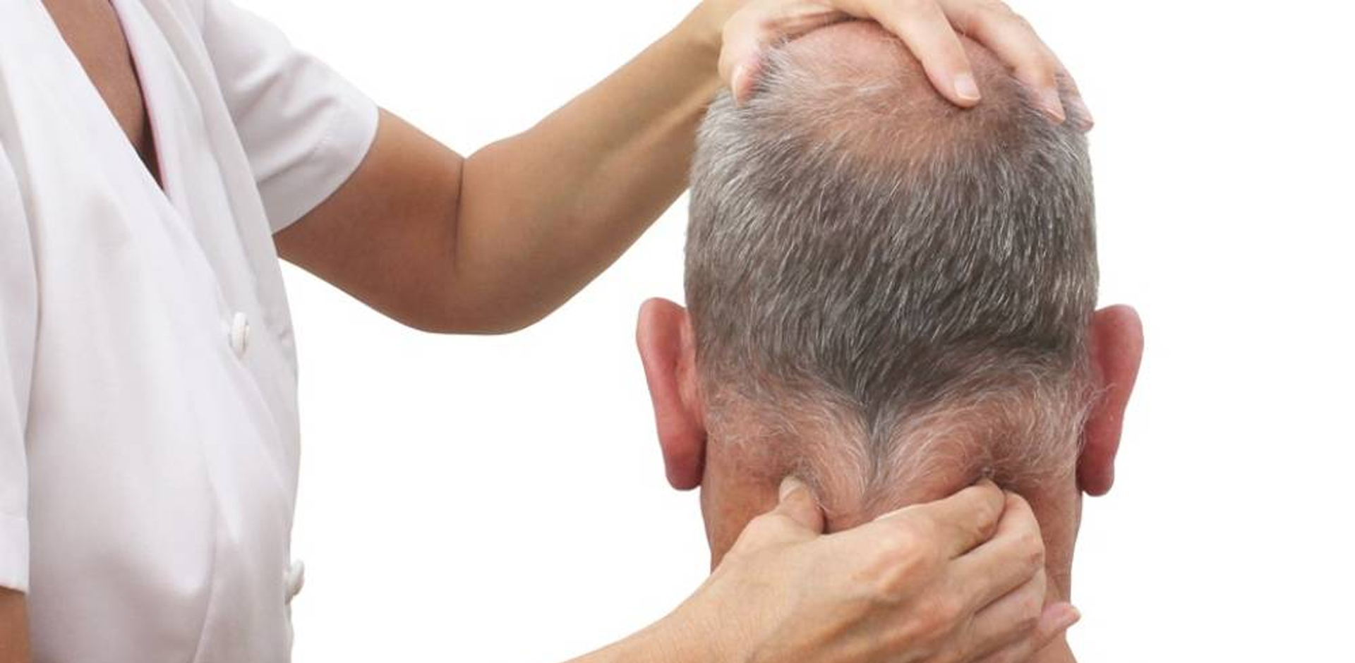
What is Neurological Physiotherapy?
Damage to your central nervous system, including your brain and spinal cord, means that the messages from your brain are not reaching the affected parts of your body. This can result in loss of movement and sensation, uncoordinated movement, weak and floppy muscles, spasms,s, and tremors.
Neurological Physiotherapy is able to kick-start the message pathways that your brain is struggling to use, to make new pathways through repetitive actions and exercises. Many of our clients who undergo Neurological Physiotherapy can improve symptoms such as, difficulties with loss of balance, loss of hand and arm, or leg and foot function, walking, spasticity, and pain.
Our team of highly trained specialist physiotherapists is experts in the treatment of all neurological conditions including:
- Stroke
- Head Injuries
- Spinal Cord Injuries
- MS
- Parkinson’s
At the first consultation, we assess and create personal goals. PhysioFunction recognizes that everybody’s goals are different. They can be anything from walking, driving to visit friends or family, returning to work, or taking up sport again. PhysioFunction has even helped a client to achieve their aim of walking down the aisle!
Physiotherapy sessions are carried out by specialist Neurological Physiotherapists, a Therapeutic Exercise Instructor, or Physiotherapy assistant, either individually or in a group exercise class. Appointments are usually within five working days. A comprehensive assessment establishes individual needs and recommendations for future treatment. Following the initial assessment, we can also advise whether there is a need to seek further assessments such as occupational therapy (available on-site at our Northampton Clinic) or speech therapies.
A stroke occurs when the blood supply to part of the brain is suddenly interrupted, or when a blood vessel in the brain bursts, spilling blood into the spaces surrounding brain cells.
Types of Stroke
There are two types of stroke:
An Ischemic Stroke is a blockage of a blood vessel supplying the brain, ultimately causing a brain infarction – or death of brain tissue. Ischemic Stroke accounts for approximately 80 % of all Stroke incidents. Ischemic Strokes can also be caused by either an abnormal narrowing or thickening of the artery wall. Also called Embolic Stroke or Thrombotic Stroke.
A Haemorrhagic Stroke is bleeding into or around the brain. Such strokes account for approximately 20 % of all strokes. Also called Intracerebral Haemorrhage or Subarachnoid Haemorrhage.
Brain cells die when they no longer receive oxygen and nutrients from the blood (Ischemic Stroke) or when they are damaged by sudden bleeding into or around the brain (Haemorrhagic Stroke). When blood flow to the brain is interrupted, some brain cells die immediately, while others remain at risk of death.
PhysioFunction’s highly specialised neurological physiotherapists are able to deliver tailored stroke rehabilitation programs.
A Stroke may affect different areas of your body. Common areas which can be enhanced through Neurological Physiotherapy include:
- Improving walking
- Improving arm and hand function after Stroke
- Reducing post-stroke shoulder pain from shoulder instability or weakness
- Enhancing mobility by addressing any foot dragging
- Reducing spasticity in limbs and muscles
- Addressing weak and spastic muscles
- Re-educating movement patterns through natural and assisted technology methods, such as:
- Functional Electrical Stimulation (FES)
- Saebo products
- Mirror therapy
Sometimes patients have been told that they have reached a point where they will no longer improve.
From our experience, we have seen improvement in clients many years after the onset of their condition. With determination and help from our specialist team of Neurological Physiotherapists, PhysioFunction can help clients regain movement and function long after they have been told they have ‘plateaued’ in their recovery.
Often called the hidden disability, people who have sustained an Acquired Brain Injury (ABI) are often faced with distinctly different challenges than people with similar impairments arising from other causes.
At PhysioFunction we work with our clients to achieve the things that matter to them. Our team understand that in order to work effectively with people who have sustained an ABI, it is essential to have a broad understanding of the physical processes of injury and recovery, the possible impact on brain functions, and the outcomes that may result – for the individual, the family and society as a whole. Therapy is an active process done with the person, not to them. Each individual’s abilities will determine how the person is able to participate.
Each therapy programme is specifically designed to meet the needs of each individual’s goals. Broadly this can be placed under the following headings:
- Motor/sensory Retraining
- Moving in bed
- Sit to Stand
- Walking
As a result of weakness or changes in tone and muscle imbalances, some individuals are at risk of developing muscle shortening and loss of joint mobility. This can then lead to more permanent reduction in function. One of the primary goals of physiotherapy intervention is to identify these muscle imbalances and provide an appropriate intervention.
Sitting here in the waiting room of the Chris Moody Centre set me thinking how had I got here? I was born in the mid-60s after what I am led to believe was a traumatic birth which resulted in my Acquired Brain Injury (ABI). As I grew up I learned the effects the ABI had on me, I found crowds of people intimidating and busy atmospheres – both visual and auditory hard to cope with. My ABI also caused me to be moody and perhaps a bit abrupt at times. My stamina is certainly reduced as is my ability to find words, I also find that I consume a lot of food and do not add weight, which I am told (from someone who had attended a lecture on nutrition for people with head injuries) is a symptom of ABI. I have epilepsy too, however this is controlled with medication.
After much frustration at fitting my square peg into a round hole (the NHS) I found myself at the Chris Moody Centre discussing a totally unrelated matter. Whilst waiting for the appointment I found a leaflet describing what PhysioFunction could provide. Almost immediately I contacted them and a few days later I found myself back at the Chris Moody Centre seeing a physiotherapist about my leg. After several sessions I mentioned how quickly I went through shoe heels – the physiotherapist talked to the Clinical Director of the company and he came back with a list of suggestions, one of which was a SAFO. So after a trip to the South Coast for a consultation which proved most fruitful and a return visit to collect the SAFO, I find that in the three years that I’ve had it I have not needed to replace my shoe heels.
At more or less the same time the physiotherapist told me about something called Neurofit, run by a personal trainer and a physiotherapist, which I likened to a gym session for people who have neurological injuries and a competitive nature. It also has improved my mental wellbeing. Initially the physiotherapist would hold my hemiplegic knee, which had a tendency to swing outwards when I did squats. After six months of Neurofit I could see how much stronger my hemiplegic knee had become.
Another chance conversation with the Clinical Director of PhysioFunction led to a visit to a surgeon who has rebalanced my wrist so that now I have a lot more use of it.
As I write this the rehab continues, what further places will I visit on this incredible journey I have no idea but it is safe to say that there will be plenty of them.
Spinal CordIinjury (SCI) occurs when the spinal cord gets damaged following an accident, falls, sporting injuries, or following diseases, such as Polio, Spinal Bifida or Transverse Myelitis.
The damage to the spinal cord might be due to severing (total or partial), crushing/compression, or over-stretching/tearing. It is not the same as having a “broken back/neck” as this refers to a fracture of one or more of the vertebrae (bones that make up the spine), which does not necessarily lead to spinal cord injury.
PhysioFunction’s Neurological Physiotherapy treatment is aimed at addressing the problems that can occur following SCI so that an individual can maximise their potential for recovery and therefore maximise their independence.
Whether the personal injury to that individual has resulted in quadriplegia/tetraplegia or paraplegia, treatment should be focused on what their needs are. Once the physiotherapist has made their assessment, and the individual has expressed what they would like to achieve, an appropriate course of physiotherapy can be agreed which will aid an overall rehabilitation and maximise recovery.
Depending on the level of Spinal Cord Injury and its nature whether it be complete or incomplete, the problems associated can include:
- Muscle weakness/paralysis
- Reduced ability to breathe
- Loss of general mobility and balance
- Loss of functional movement
- Poor posture
- Pain
Treatment should be focused upon that individual and tailored specifically to their condition. A treatment programme is formulated following a thorough physical assessment which might include:
- Stretching activities to maintain muscle and tendon length and reduce or keep muscle spasms/spasticity to a minimum.
- Flexibility and strengthening exercises for the whole body.
- Breathing exercises to maximise lung function and prevent chest infection.
- Balance and posture exercises which can help to reduce pain associated with poor posture and balance impairment and ensure correct transfer techniques (in/out of wheelchair, bed, toilet/bath, car etc.)
- Functional activities to improve fundamental movement patterns such as rolling over and sitting up, and standing where appropriate.
- Walking re-education, if there is sufficient muscle activity and power in the legs.
- Your physiotherapist might also be able to advise an individual on use of appropriate equipment such as wheel-chairs and pressure releasing cushions, exercise equipment and electrical muscle stimulators (EMS)/Functional Electrical Stimulation (FES) e.g. Odstock PACE, WalkAide, Bioness, Ottobock MyGait, Ottobock ActiGait, and Exoskeletons including REX.
- With appropriate treatment and by challenging an individual during recovery, together with sound advice and encouragement, Neurological Physiotherapy can indeed maximise your independence.
Neurological Physiotherapy in Multiple Sclerosis addresses a number of key areas which can dramatically change a person’s ability to do day to day activities and maximise their independence in all aspects of life, including leisure and vocational activities
The first part of Multiple Sclerosis treatment is to improve posture. This means putting the right muscle in the right place, at the right time, so that it can work most efficiently. This is essential for balance, walking and arm and hand function.
Neurological Physiotherapy can help re-educate poor balance, even if there is no specific muscle weakness, Multiple Sclerosis can alter the way in which we move, making you feel as if you are more likely to fall.
If you have difficulties with your balance, it is likely that your walking is a challenge as well. People with Multiple Sclerosis often have problems with tripping. There are a number of reasons why walking maybe affected in Multiple Sclerosis. It may be general or specific muscle weakness. The general weakness can be addressed by various types of exercise. If it is a more specific weakness the treatment is likely to be more specialised using rehabilitation technology. The most common of these is the treatment for drop foot or a floppy foot with Functional Electrical Stimulation (FES).
Hand and limb function can also be helped with physiotherapy or some of the rehabilitation technology available at PhysioFunction.
Many people with MS suffer with muscle spasms, pain or stiffness in the muscles which we call spasticity. The physiotherapist can help reduce the problems associated with this type of altered muscle function and leg weakness. We may use a number of different treatment approaches including rehabilitation exercises, the use of splints or orthoses or working with the doctors to help with the prescription of medication.
Lastly, many people with Multiple Sclerosis suffer from fatigue. This can be the biggest limiting factor when trying to do every day activities. Fatigue is a symptom of MS, however people also get fatigued because of the extra effort they have to invest in all of their activities. Neurological Physiotherapy will work with you to help you manage the fatigue so you can save your energy for the good stuff!
What Is Bell's Palsy
Bell’s palsy is a condition in which the muscles on one side of your face become weak or paralyzed. It affects only one side of the face at a time, causing it to droop or become stiff on that side.
It’s caused by some kind of trauma to the seventh cranial nerve. This is also called the “facial nerve.” Bell’s palsy can happen to anyone. But it seems to occur more often in people who have diabetes or are recovering from viral infections.
If it happens to you, you may fear you’re having a stroke. You’re probably not. A stroke that affects your facial muscles would cause muscle weakness in other parts of your body, too.
What Causes It?
Most doctors believe that it’s due to damage to the facial nerve, which causes swelling. This nerve passes through a narrow, bony area within the skull. When the nerve swells — even a little bit — it pushes against the skull’s hard surface. This affects how well the nerve works.
Bell’s Palsy: How Is It Diagnosed and Treated?
There’s no test that can tell you for sure if you have Bell’s palsy. In fact, doctors usually find out through what they call a “diagnosis of exclusion.” That means in most cases, they determine you have Bell’s palsy only after other conditions have been ruled out.
Your doctor will start by doing a complete and careful physical exam. If he suspects you have Bell’s palsy, he’ll try to close your eyelid on the affected side of your face. If it doesn’t close, it’ll signal that you have what doctors call “the Bell phenomenon.” With this condition, your eye rolls upward and outward when you try to close it.
Your doctor will then try to rule out other conditions. He’ll probably test your hearing and sense of balance. He may also order several tests, such as skull X-rays, a computed tomography (CT) scan, or magnetic resonance imaging (MRI). Electrical testing may help clarify the diagnosis. It may also help him predict how fast and fully you’ll recover.
What Are the Treatments for Bell’s Palsy?
There aren’t any that can stop it. If your doctor suggests your symptoms might be triggered by the herpes virus (herpes simplex 1) or by shingles (herpes zoster), he may give you an antiviral medication, like acyclovir. But there’s no research to show these medications work to reduce Bell’s palsy symptoms.
Your doctor may also give you a short course of corticosteroids (like prednisone). The goal is to decrease swelling of your facial nerve. This may shorten the duration of your Bell’s palsy symptoms.
In the meantime, your doctor will tell you to take extra care to protect your eye on the affected side. He may suggest you wear an eye patch since you won’t be able to blink. If your eyes are tearing less than normal, you may have to use eye drops to keep them from drying out.
What is Chronic Pain?
Chronic Pain is any pain that lasts longer than 3 months. This means that the pain you are feeling is not because something is injured, it is simply because the brain thinks it is normal to feel pain. The brain is always learning new things. If you repeat something often enough, it will begin to make a pattern so that the brain can reproduce it without thinking. For example, when you walk you don’t always have to think about how to do that. Your brain has a pattern to follow so that it can happen automatically.
The same happens with the pain signal. If you are always triggering your pain signal, your brain will learn that those activities should always be painful. Overtime, if you trigger your pain repeatedly, it will create a pattern that the brain will continue to use even after your injury has healed. This is how people continue to feel pain from an injury several months, and even years, after the original injury occurred.
Types of Treatments:-
Physiotherapy:
Science is only beginning to understand chronic pain and the variety of effects it has on the body. Although there are still many unknown factors about chronic pain, we know enough to help you manage your pain, reduce it and sometimes even get rid of it completely. Physiotherapists are movement experts and we can help you identify if the pain you feel is justified, or if you have chronic pain from an old injury. From here, we can provide treatment and share strategies for you to manage your pain at home. It is never too early or too late to start training your brain to move without pain and get back to a normal life.
Neurology:
The cause of chronic pain is not always obvious. Some people might be told that “it’s all in your head” and that “there’s nothing wrong”. They will often go through multiple tests like X-Rays or MRIs without finding a reason for the pain. This does not mean that the pain is not real. The pain that you are feeling is very real.
A physiotherapist can examine your movements and check for any possible causes of the pain. They can verify if any nerves are being compressed or irritated. They can check to see if any ligaments are injured. These are all things that could cause pain, but can’t be seen on an X-Ray or MRI. If there is a cause, a physiotherapist can give you specific recommendations on how to promote healing.
If there is no cause, then we can determine that you are feeling chronic pain. If this is the case, a physiotherapist with experience in neurology can help you retrain your brain to move without pain. We do this by working within a pain-free range to start. The goal is to teach your brain that the movements are not supposed to be painful. You should feel some relief after the first few treatments. Your physiotherapist should show you some exercises to make sure that the pain keeps improving, and doesn’t come back!
Exercises:
When choosing exercises for your chronic pain, you want to make sure that you stay within a pain-free range. If you’ve been feeling pain for a long time, you might be surprised at how little you can do before triggering your pain. It is very important to not push this barrier during your exercises. Remember, you are trying to teach your brain that movements are not painful.
Depending on where you feel your pain, a physiotherapist can prescribe you specific exercises so that you can target the movements that are usually the most painful for you. They will help you progress until you feel comfortable with your progress. Then you can gradually find things that are more challenging, to get back to your daily activities without pain
The type of exercise that you do is important. Your brain is most likely to remember how to move without pain if you practice during activities that are meaningful to you. We want to make the changes permanent, so we use activities that you enjoy in the clinic, and in your Home Exercise Program.
Frequency:
In order to make the brain understand that movements are not painful, it is important to practice your exercises correctly, and often. Just like remembering a new phone number. You might choose to review it, to make sure you remember it correctly, then you will repeat it to yourself often to make sure that you don’t forget it.
The same strategy applies to your movements. You need to make sure that you are moving without pain every time you perform an activity, like walking, or climbing stairs. For this reason, our treatment plans are designed with visits 1 to 2 times per week.
Duration:
The length of treatment has two components. First, the type of session, and second, the total length of the treatment plan. There are two types of sessions to choose from: regular treatment and complex treatment. Most people with chronic pain benefit from regular treatment sessions, that focus on only one problem. If you have pain in more than one place, sometimes a complex treatment session is beneficial to address more than one problem at once.
For your first treatment plan, we often suggest a duration of 4 weeks. This allows us to see how you progress and how you are doing with your Home Exercise Program. After these 4 weeks, you may choose to continue on your own, or to continue for another treatment plan, which can be for up to 12 weeks.
The duration of your treatment will be discussed with your physiotherapist on the day of your initial assessment. Depending on what the findings are, and what your goals are, you will talk about what type of treatment session is most appropriate and how many weeks you should start with.
What is Epilepsy?
Epilepsy is a condition that causes children and adults to have repeated seizures. Seizures result from abnormal stimulation of brain cells known as neurones which causes the body to react in an unusual way.
The severity of the seizures can vary from person to person, some seizures can cause people to loose awareness or freeze for a couple of minutes others can cause convulsions (uncontrollable shaking) and loss of consciousness.
Epilepsy often starts in childhood due to an unknown reason, others can be a result of brain injury, eg a severe head injury.
Physiotherapy Treatment for Epilepsy
Physiotherapy treatment is usually for secondary complications from falls such as broken bones or a head injury.
Treatment can include::
- • Education is the main treatment, to help prevent injury. For example, padding out sharp corners in the house to prevent damage if fallen on. Advice on special swimming equipment to prevent drowning in seizures that happens in the pool.
- • Treatment for broken bones will be after the fracture site has completely healed or is classed as stable (this is usually from open reduction and internal fixation). Upper limb usually takes 6-8 weeks and lower limb can take 8-12 weeks. Physiotherapy treatment after this is usually consists of strengthening muscles that have got weaker from being immobilised and stretching muscles that are stiff from being in the cast for such a long period of time.
What is autism?
Autism is a lifelong developmental disorder. Autism affects the way a person relates to the world around them; to the autistic individual, the world seems a mass of people, places, and objects that all seem to be able to interact intuitively (by themselves with no prompts or teaching), autism suffers find this somewhat difficult.
Autism is not a learning difficulty as such, but most autistic children present with learning difficulties.
Autistic disorders fall into what’s known as an autistic spectrum, within this spectrum all the disorders are based on three key difficulties:
- Difficulty with communication
- Difficulty with social interaction
- Difficulty with social imagination
High-functioning Autism forms part of the autistic spectrum alongside Asperger’s. Although they present similar difficulties to the patient, they sit at opposite ends of the spectrum:
- Asperger’s – a relatively mild form of the disorder characterised by communicative difficulties
- High-functioning autism – a disorder characterised by significant language development.
- High-functioning autism affects speech, understanding and communicative abilities.
What are the symptoms of autism?
No two people with autism will present with the same symptoms to the same degrees, every person will have a unique set of difficulties they experience within everyday life
In general, autistic children tend to be quiet individuals who like to keep themselves to themselves; they often avoid eye contact as they prefer to stay out of conversations as opposed to getting involved. This is also characteristic due to the communication difficulties, and the anxiety if they do say something, it will be the wrong thing.
Many children with autism also have associated physical limitations, may lag behind in the achieving of developmental milestones, sometimes these difficulties are missed by parents, as they struggle to engage.
How will physiotherapy help individuals with autism?
Physiotherapy will consist of an individualised treatment programme to match the needs of the individual. It will aim to address difficulties faced in the individual’s everyday life, and seek to achieve any other goals. Physiotherapy is an interactive process that often takes time and trust for the child to engage, once completed, it is hoped the programme will have a positive impact both on physical ability and psychological wellbeing.
The physiotherapy intervention may include:
- Practice of basic movement skills, such as sitting, standing and playing
- Practice of more complex skills such as kicking, throwing and catching- these are important for physical development, but also for social engagement in sports, recess and general play.
- Techniques for helping build muscle strength, coordination and skills
- Building social/physical skills
What are the benefits of physiotherapy for people with autism?
Physiotherapy for people with autism has a number of benefits, both physical and psychological:
- Increased self confidence
- Improved interaction skills
- Improved physical skills
- Improved quality of life
What is Neuropathy?
The word Neuropathy is derived from two words neuron and pathology. Neuron means- nerve cell(a cell that is specialized to conduct nerve impulses). Pathology means- any deviation from a healthy or normal condition.
On clinical context Neuropathy is commonly refered to as Peripheral Neuropathy. The term peripheral neuropathy designates a disturbance in function of one or more peripheral nerves. Several types of peripheral neuropathy are distinguishable by the extent of involvement. Depending upon the underlying cause, there may be selective involvement of motor, sensory, or autonomic fibers or more diffuse involvement of all fibers in the peripheral nerve.
Symptoms of Peripheral Neuropathy:
Peripheral Neuropathy produces symptoms depending upon affected nerve. They can be sensory, autonomic or motor changes. The main symptom is the numb feet with tingling and can also cause weakness, coordination problems, pain, burning and invisible ‘glove-like’ sensation and abnormal heart rate, reduced sweating and sexual problems.
Classification of Peripheral Neuropathy:
Peripheral neuropathy can be classified into more than 100 forms and produces different set of symptoms and have different prognosis. Motor, sensory and autonomic nerves damage can occur. If the motor nerves are affected then imbalance occurs in the coordination of walking, holding things and other voluntary muscle movements. Damage in sensory nerves results in the loss of sensations like touching or pain whereas autonomic nerves affect the involuntary nerves controlling vital organs.
Four types depending upon number of nerve damage and the nerve cells are autonomic neuropathy, mononeuropathy, mononeuritis multiplex and polyneuropathy. When only one nerve is affected then it is called as mononeuropathy. It can be caused by compression to the nerve, carpel tunnel syndrome or some infection and nerve inflammation causing tingling feet.
MONONEURITIS:
Mononeuritis multiplex occurs when multiple nerves are damaged here and there in the body due to diabetes mellitus (diabetic neuropathy), Churg-Strauss syndrome, HIV, amyloidosis and rheumatoid arthritis. It is present with dull pain in legs and back mostly at night. Diabetics may have severe pain in thigh of either side with weakness and knee reflex absence.
POLYNEUROPATHY:
Polyneuropathy affects nerve cells anywhere in the body irrespective of the nerve path. It can cause changes in axon, neurons cell bodies and myelin sheath surrounding axons. Distal axonopathy is the condition affecting only the axons with intact neurons. In sensory neuronopathy and motor neuron disease sensory and motor neurons are affected respectively. Polyneuropathy attacks organs on either side. It produces symptoms such as numb feet, burning, erectile dysfunction and imbalance in bladder function. This neuropathy treatment involves three steps. It starts with removing the cause, then strengthening muscles and their function and in the last pain relief by using neuropathy creams containing capsaicin.
AUTONOMIC NEUROPATHY:
The fourth pattern in the peripheral type of neuropathy is the autonomic neuropathy causing alterations in the autonomic nervous system. It affects the non-involuntary nerves reaching urinary bladder, digestive system, sexual organs and heart. Chronic diabetes patients are more prone to this neuropathy. Autonomic neuropathy is present in combination with other neuropathies. It produces symptoms such as incontinence of urine, pain in abdomen with vomiting, diarrhea or constipation, tachycardia, hypotension and impotency.
Common Causes Of Neuropathy:
It’s not always easy to pinpoint the cause of peripheral neuropathy, because a number of factors can cause neuropathies. These factors include:
Alcoholism :Many alcoholics develop peripheral neuropathy because they make poor dietary choices, leading to vitamin deficiencies.
Autoimmune diseases :These include lupus, rheumatoid arthritis and Guillain-Barre syndrome.
Diabetes :When damage occurs to several nerves, the cause frequently is diabetes. At least half of all people with diabetes develop some type of neuropathy.
Infections :Certain viral or bacterial infections can cause peripheral neuropathy, including Lyme disease, shingles (varicella-zoster), Epstein-Barr, hepatitis C and HIV/AIDS.
Exposure to poisons :These may include some toxic substances, such as heavy metals, and certain medications – especially those used to treat cancer (chemotherapy).
Nherited disorders :Examples include Charcot-Marie-Tooth disease and amyloid polyneuropathy
Trauma or pressure on the nerve :Traumas, such as motor vehicle accidents, falls or sports injuries, can sever or damage peripheral nerves. Nerve pressure can result from using a cast or crutches, spending a long time in an unnatural position or repeating a motion many times — such as typing.
Tumors :Growths can form directly on the nerves themselves, or tumors can exert pressure on surrounding nerves. Both cancerous (malignant) and noncancerous (benign) tumors can contribute to peripheral neuropathy.
Vitamin deficiencies :deficiencies B vitamins — B-1, B-6 and B-12 — are particularly important to nerve health. Vitamin E and niacin also are crucial to nerve health.
Other diseases :Kidney disease, liver disease and an underactive thyroid (hypothyroidism) also can cause peripheral neuropathy.
Treatments for Peripheral Neuropathy:
Effective prognosis and treatment of peripheral neuropathies relies heavily on the origin of the nerve damage. For example, peripheral neuropathies caused by vitamin deficiencies can often be halted — even reversed — with vitamin therapy and an improved diet. Likewise, nerve damage brought on by alcohol abuse can often be improved by avoiding alcohol. Peripheral neuropathies caused by toxic substances or medications can often be corrected in much the same way. When neuropathy is related to diabetes, careful monitoring of blood sugar levels may slow its progression and curb symptoms.
Early diagnosis and treatment of peripheral neuropathy is important, because the peripheral nerves have a limited capacity to regenerate, and treatment may only stop the progression — not reverse damage. If you have become severely impaired, you may need physical therapy to help retain strength and avoid muscle cramping and spasms.
Surgical treatment may be recommended for people with nerve damage from injury or nerve compression. Mobility aids, such as a cane, walker, or wheelchair, may be helpful. For pain, your doctor may prescribe pain medication.
Encephalitis
Encephalitis is the medical term used to describe severe inflammation of the brain tissue. Encephalitis is either caused by a bacterial or viral infection.
What is encephalitis?
Encephalitis is inflammation of the brain tissue. Encephalitis is either caused by a bacterial or a viral infection, or an irregular autoimmune response in which the bodys own immune system attacks the brain tissue causing it to become inflamed.
In most circumstances the brain is effectively protected from infection by a thick membrane, which is known as the blood brain barrier. However in a minority of people an infection can penetrate the blood-brain barrier. The infection can then enter brain cells and nerve cells causing them to become inflamed. Inflammation causes the brain cells and nerve cells to become damaged, resulting in a loss of normal brain function.
Encephalitis can permanently damage the brain tissues, resulting in an acquired brain injury. If encephalitis is not medically treated as soon as possible then it can lead to a coma and eventually death.
Effects of encephalitis
Initially encephalitis presents itself by way of flu like symptoms such as a high temperature, headaches, stiffness and fatigue. However the headache you experience may become more severe and you may begin to vomit. Instances in which your body temperature is extremely high often result in confusion, drowsiness, and seizures.
Physiotherapy treatment for encephalitis:
In order to maximise your recovery following encephalitis it is important that physiotherapy treatment is initiated as soon as possible in order to maintain and improve the current level of functional ability you possess. At Liverpool Neuro Physio your physiotherapist will:
- Provide assistance in task practice. Your physiotherapist can teach you simple techniques to break down a task into simple steps in order to perform it effectively.
- Provide gait training in order to improve the coordination of your limbs during walking.
- Provide strengthening exercises to maintain muscle strength and minimise the occurrence of muscle wastage, which is often a secondary complication of inactivity
- If appropriate, provide advice on additional equipment to increase your functional independence.
The long term complications experienced as a result of encephalitis are very inhibiting and they often make it difficult to carry out simple everyday tasks. At Liverpool Neuro Physio our experienced neurological physiotherapists can provide you with the advice and treatments you need in order to cope with your condition and maintain your functional independence in order to make your life as fulfilled as possible.
Benefits of physiotherapy treatment for encephalitis
Benefits of physiotherapy treatment for the long term motor complications experienced as a result of Encephalitis may include:
- Improved coordination
- Improved balance
- Improved muscle control
- Reduced anxiety
- Improved confidence
- Improved independence
- Improved quality of life
At Liverpool Neuro Physio our experienced physiotherapists aim to improve your functional abilities where possible by providing you with effective treatments to manage the long term complications you may experience following encephalitis.
What Causes Lumbar Degenerative Disc Disease?
Although the cause of lumbar degenerative disc disease is unknown, it seems to be associated with aging as your intervertebral discs degenerate naturally due to regular wear and tear.
Risk factors that increase your likelihood of developing it include
- Increasing age
- Trauma or injury to your back
- Repetitive strain to your back, such as a job requiring frequent heavy lifting
- Being overweight
- Smoking
- Prolonged sitting and inactivity
- Poor posture
- Musculoskeletal imbalance, such as scoliosis
What are the Signs and Symptoms of Lumbar Degenerative Disc Disease?
Everyone experiences some level of degeneration in their intervertebral discs over time, but not everyone will experience symptoms.
If they do, symptoms of lumbar degenerative disc disease typically include:
- Dull pain in the neck
- Stabbing pain after strenuous activity or strain on the neck
- Increased pain with movement, such as turning or bending your neck down
- Difficulty moving and decreased range of motion
- Muscle tension or spasms
- A sharp, stabbing pain or weakness and tingling that radiates down into the shoulders, arms or hands, known as cervical radiculopathy
How is Lumbar Degenerative Disc Disease Treated?
Mild lumbar degenerative disc disease can be treated at home or in a clinical setting
To get pain under control, you can:
- Rest your back
- Apply heat and cold therapy
- Avoid activities that cause pain or put stress on your back
- If needed, take anti-inflammatory painkillers such as Advil or Aleve
Moderate to severe levels of lumbar degenerative disc disease should be treated in a clinical setting.
Physiotherapy for Lumbar Degenerative Disc Disease:
Physiotherapy is a drug-free and non-surgical treatment that focuses on reducing pain, regaining strength, and increasing joint mobility, function, and quality of life.
At pt Health, you’ll receive a thorough assessment which addresses the source of your problem.
Depending on the cause and severity of your lumbar degenerative disc disease, physiotherapy treatment can include::
- Strengthening and range of motion exercises
- Personalized exercise plan
- Active stretching
- Functional retraining and activity modification
- Patient education
- Cross-disciplinary pain-relieving therapies such as:
- Interferential current therapy (IFC) or TENS therapy
- Therapeutic ultrasound
- Manual therapy (joint and soft tissue mobilizations)
- Heat and cold therapy
- Occupational therapy
What is motor neurone disease?
Motor neurone disease (MND) is the name given to a group of disorders that affect nerve cells in the brain and spinal cord leading to muscle weakness, wasting and loss of function. It also affects the muscles that enable us to speak, breathe and swallow making these processes difficult. Motor neurone disease is a rare progressive neurological condition that commonly affects people between the age of 50 and 70. Over time the symptoms of MND become worse.
Physiotherapy treatment will promote functional independence by maintaining muscle strength and length and increasing energy levels and sense of well-being. Physiotherapy will also improve a person’s ability with everyday tasks and improve their quality of life.
Types of motor neurone disease
There are three main types of MND:
Amyotrophic lateral sclerosis (ALS)) is the most common type of MND and causes muscle weakness and stiffness.
Progressive bulbar palsy (PBP) :accounts for 20% of MND cases and causes difficulty with speaking and swallowing.
Progressive muscular atrophy (PMA) :accounts for 10% of cases and causes muscle weakness, wasting and twitching.
MND affects people in different ways and there is a difference in how quickly each type of MND progresses.
What causes motor neurone disease?
Motor neurone disease (MND) is caused by progressive damage to the motor neurones in the brain and spinal cord. Motor neurones send messages from your brain to your muscles allowing you to move. They also help regulate many of the body’s automatic processes, such as breathing and swallowing.
It is still unclear how the motor neurones become damaged in MND.
What are the effects/symptoms of motor neurone disease?
The symptoms of MND depend on the type of MND you have. The main symptom of MND is muscle weakness that becomes progressively worse over time. The first symptoms commonly develop in the hands and arms, or feet and legs. Although physical functions decline, cognitive function remains intact and your memory, sight and smell remain unchanged. Other initial difficulties may include some of the following:
- Stumbling
- Dragging of a foot
- Difficulty gripping objects
- Stiff joints
- Fatigue especially with daily activities
- Pain and discomfort
- Muscle wasting
- Spasticity or stiffness in the arms and legs.
- Difficulties with speech, chewing and swallowing.
- Breathing problems
As MND progresses, a person with MND may become more almost totally immobile requiring them to use a wheel chair to complete everyday tasks. The speed of the progression varies greatly with individuals.
Furthermore, people with MND may experience a ‘plateau’ for weeks or even months where symptoms remain the same and no deterioration occurs.
Physiotherapy treatment is very important and should be started as soon as possible. Physiotherapy will help improve balance, posture and mobility. It will also make movement more functional and reduce the impact the physical symptoms have on your life.
Physiotherapy for motor neurone disease
At Physio.co.uk we understand that being told that you have motor neurone disease (MND) can be an emotionally experience and the news can be difficult to take in. Physio.co.uk recommend an early intervention which is very important as this allows time for an effective relationship to be built up with the physiotherapist which will become invaluable as your condition progresses. Our dedicated physiotherapists at Physio.co.uk will promote functional independence by maximising your potential and increasing your self-esteem.
At Physio.co.uk our specialised neurological physiotherapists acknowledge that MND affects everyone differently so treatment is tailored to your needs. An initial assessment will look at how MND affects you and goals will be developed centred around you and those close to you. Physiotherapy treatment will be focused on:
- Minimising abnormalities of muscle tone
- Maintaining muscle strength for as long as possible
- Preventing secondary problems such as soft tissue contractures and infection
- Advice on postural management
- Facilitating the use of efficient functional movement patterns including the quality of walking.
- Helping stiff muscles and joints
- Increasing range of motion
- Increasing flexibility
- Improving balance and posture
- Increasing comfort when sitting, standing or sleeping
- Reducing the risk of falling
- Education and advice about MND
- Increasing energy levels
- Improving sense of well being
- Helping maintain independence
Assessment of your individual needs will be taken on a regular basis as your physical symptoms will change over time. Physiotherapy treatment for motor neurone disease includes:
- Exercises to maintain muscle strength and length and how to incorporate these into everyday activities.
- Teach techniques that help make some automatic movements and functional abilities easier. For example, walking, sitting down and standing up are some of the tasks that may become difficult as MND progresses, but can be improved by learning new ways of doing things.
- Balance training to help improve confidence and reduce the risk of falling.
- Teaching and advising family and carers on positioning, transfers and mobility aids.
- Gentle exercise and massage will help reduce your anxiety
- Hydrotherapy treatment will also help relax and stretch tight muscles and help with mobility.
What is Myasthenia gravis?
Myasthenia gravis or ‘’grave muscle weakness’’ is an autoimmune neuromuscular condition affecting the muscles in the body causing varying degrees of weakness. Myasthenia gravis is caused by the body’s immune system attacking the nerves supplying the muscles. Myasthenia gravis usually affects younger women, and men over the age of 60.
What causes Myasthenia gravis?
It has been suggested the body’s own immune system releases antibodies that block important neurotransmitters. These neurotransmitters make your muscles contract. This damage therefore impairs the nerve supplying the muscle, so the muscles do not contract well and become weak and tired.
It is unclear what causes the immune system to do this. Some people with Myasthenia gravis find that tiredness and stress can bring on symptoms but others find their symptoms disappear spontaneously and don’t return for many years.
What are the effect / symptoms of Myasthenia gravis?
Myasthenia gravis affects the voluntary muscles of the face, eye, neck and jaw arms and legs. The most noticeable symptom is drooping of the eyelid making a person look tired. A person with myasthenia gravis will also have difficulty controlling eye movement, eating and swallowing and weakness in arms, hands, fingers and legs. The muscles that control breathing may also be affected causing problems with breathing.
Physiotherapy for Myasthenia gravis?
Neurological Physiotherapy treatment will help a person with myasthenia gravis. Our specialised neurological physiotherapists at Physio.co.uk understand that Myasthenia gravis can have a considerable impact you both physically and emotionally. Physiotherapy will help improve your energy levels and sense of well-being. The exercise program will depend on your individual needs and will be centred around:
- • Increasing fitness and energy levels
- • Increasing muscle strength
- • Improving quality of sleep
- • Support with everyday tasks
- • Advice on diet
- • Improving posture when sitting, standing and sleeping
- • Increased ability to relax
- • Advice on supportive devices
- • Improving quality of life
It is important to plan your activities to coincide with the time when your energy levels are at their highest. Our specialised neurological physiotherapists will help keep your work, home and social life as active as possible. Physiotherapy treatment will include:
- • Muscle strength training to improve weakness in the muscles
- • Exercises to enhance functional abilities
- • Breathing control and assisted coughing if appropriate to maintain a clear chest.
- • Exercise to increase stamina and reduce fatigue
- • Relaxation therapy and massage to ease sore muscles.
- • Hydrotherapy
What is neurofibromatosis?
Neurofibromatosis (NF) is a genetic disorder which affects the central nervous system (brain and spinal cord) and the skin and causes non-cancerous tumors (i.e., neurofibromas) to grow on nerve tissue. Symptoms of NF depend on the type (Nf1 or Nf2) number, size and location of the tumour. Neurofibromatosis can be inherited or occur spontaneously.
Types of neurofibromatosis
There are two main types of neurofibromatosis which include
- Nf1 – also known as von Recklinghausen’s disease or peripheral
- Neurofibromatosis.
- Nf2 – also known as bilateral neurofibromatosis.
Diagnosis of neurofibromatosis
A diagnosis of neurofibromatosis can be made be taking a detailed family history, physical examination for clinical signs of the disease (for example skin lesions), and magnetic resonance imaging (MRI). Genetic testing may also be used to confirm a diagnosis.
What are the effects/symptoms of neurofibromatosis?
The symptoms of NF vary from person to person. Some people will be mildly affected with very few health problems whilst others will have some serious health problems, making everyday tasks difficult. The two different types of neurofibromatos have different symptoms:
Symptoms of Nf1 include abnormal skin pigmentation and lumps under the skin. However complications associated with this type of neurofibromatosis can be serious and include:
- Scoliosis (curvature of the spine),
- Bone problems that are present at birth or from an early age, such as bowing of the legs below the knees
- Coordination and memory problems
- Problems with speech, sight and hearing
- Increased risk of epilepsy
Symptoms of Nf2 are more serious and include acoustic neuroma (tumours in the ear) and cataracts (cloudy patches over the lens in the eye). However, secondary problems may include:
- Tumours on the skin.
- Brain tumours – which are usually benign, but can put pressure on the brain causing headaches, vision problems, difficulty balancing and epilepsy
- Spinal tumours or nerves surrounding it can cause symptoms such as muscle weakness, abnormal sensation and pain. Spinal tumours around the neck can affect the face (usually only on one side), making it hard to blink, smile or swallow.
Physiotherapy treatment will target your child’s physical symptoms and maximise their potential with daily activities.
Physiotherapy treatment for NF
Physiotherapy treatment will depend on the type of neurofibromatosis your child has and their individual symptoms. At Physio.co.uk, our specialised neurological physiotherapists will tailor the initial assessment and following treatment to your childs symptoms and needs. Physiotherapy treatment for neurofibromatosis will help:
- Increase muscle strength
- Prevent soft tissue shortening
- Improve posture
- Increase balance
- Improve mobility
- Improve independence with everyday tasks
- Reduce pain and increase comfort
Our neurological physiotherapists understand that neurofibromatosis effects every child differently. Your physiotherapist will support your child through a rehabilitation programme of functional activities and exercise to maximise your childs recovery and improve their quality of life. Physiotherapy treatment may include:
- • Structured exercise programme to strengthen muscles and reduce muscle wasting
- • Muscle stretching to help lengthen tight muscles and improve range of movement
- • Exercises to build stamina and reduce fatigue
- • Correcting and varying posture in different positions to improve balance and coordination
- • Advice on orthotic devices and walking aids if appropriate
- • Hydrotherapy treatment to strengthen and stretch specific muscles in a relaxed environment.
Erbs palsy
What is Erb’s palsy?
Erb’s palsy is also called Erb-Duchenne palsy refers to paralysis of the upper brachial plexus caused by injury to the superior roots of the brachial plexus (C5–C6). The position of the limb, under such conditions, is characteristic: the arm hangs by the side and is rotated medially; the forearm is extended and pronated (see Figure 4). The arm cannot be raised from the side; all power of flexion of the elbow is lost, as is also supination of the forearm — “waiter’s tip hand position” (hand turning backwards). Moro reflex is absent. Meanwhile, the grasp reflex is present which differentiates it from another brachial nerve palsy, Klumpke palsy (C7, C8, and T1). The newborn baby may have impaired sensation on the outer side of the arm and/or phrenic nerve palsy (C3, C4, and C5a).
Erb’s palsy relates to an injury to the superior roots of the brachial plexus of C5 and C6. Traction of the shoulder can injure the brachial plexus (particularly C5 and C6) during delivery. Erb’s palsy usually occurs in macrosomia (large baby) and shoulder dystocia infants who needed to be pulled from the birth canal 1). This maneuver causes stretching of the brachial plexus which may injure it. Other causes that lead to forced pulling of the shoulder include breech delivery, multipara mother, maternal obesity, maternal diabetes, or vacuum and forceps delivery 2). Erb’s palsy also may occur in cesarean section 3). Erb’s palsy is a common birth injury with estimated prevalence ranges of 0.2 to 2.0 of every 1000 births 4). Erb’s palsy causes significant arm weakness that affects 0.4 to 5 in 10,000 births.
The resulting traction injury may vary from neurapraxia (a temporary loss of motor and sensory function due to blockage of nerve conduction) or axonotmesis (a more severe injury where the nerve axons and their myelin sheath are damaged but the endoneurium, perineurium and epineurium remain intact) to neurotmesis (the most serious nerve injury both the nerve and the nerve sheath are disrupted. While partial recovery may occur, complete recovery is impossible) and avulsion of rootlets (most severe form of brachial plexus injury where the nerve roots are torn off the spinal cord) from the spinal cord. Most individuals with neuropraxia injuries recover spontaneously with a 90-100% return of function. Axonometric lesions involve disruption of axon and myelin sheath. Recovery of axonometric lesions depend on the level of lesion, and it may take months to heal with proper treatment including physiotherapy.
Neurotmesis is the worst prognosis, as the lesion causes irreversible damage affecting the axon, myelin sheath as well as the supporting structures through the nerve. The proximal end of nerve tries to regenerate by forming a neuroma in which the nerve has torn and healed but scar tissue puts pressure on the injured nerve and prevents it from conducting signals to the muscles. A nerve that is completely pulled apart cannot repair itself, so the muscles it controls are paralyzed. Sometimes it is possible to have an operation to mend the nerve and restore some function to the muscle, if this is necessary, the child is likely to be left with some residual weakness in the arm. In rare cases the nerve may be torn away from the spinal column itself – this is called an AVULSION in these cases it is imperative to consult a specialist in Brachial Plexus Injuries as soon as possible to discuss a treatment plan.
Possible complications include:
- Abnormal muscle contractions (contractures) or tightening of the muscles. These may be permanent.
- Permanent, partial, or total loss of function of the affected nerves, causing paralysis of the arm or arm weakness
The treatment of Erb’s palsy depends on the nature of damage, which is either nerve bruise (neuropraxia) or nerve tear. Nerve bruise (neuropraxia) usually resolve on its own over a period of months. However, in the latter case i.e. nerve tear, the following multiple approaches are advised, physiotherapy for regaining muscle usage 5), surgical interference 6) of nerve transplants (usually from the opposite leg), subscapularis releases, latissimus dorsi tendon transfers and rehabilitation therapy.
Fortunately, nearly all affected infants with Erb’s recover in the first few months 7). Erb’s palsy resolves completely in the first year of life in 80% to 96% of patients and in nearly 100% if recovery begins within four weeks of birth 8). Rarely is a child left with a significant disability. Hydrotherapy is a form of physical therapy used because of the anti-gravity environment. It minimized the stress on the musculoskeletal frame, allowing the neonate to move with less pain and at the same time strengthening muscles and reducing spasms. Paralyzed muscles relax in the opposite position of the waiter’s tip posture by abduction at the shoulder, external rotation of the arm, and supination of the forearm. Physiotherapy begins after two weeks. Surgical intervention, nerve graft, or nerve decompression is the next step if there is no response after 3 to 6 months 9).
Denervated muscle changes begin at the moment of injury, and without reinnervation, becomes irreversible at 18 to 24 months. These time-sensitive, irreversible changes are the scientific basis for early management recommendations. Neglected cases may lead to 20% to 30% permanent deficit of the nerve function
Erb’s palsy causes
Erb’s palsy results from a lesion at Erb’s point where C5 and C6 unite to form the upper trunk of the brachial plexus.
Damage to the upper nerves that make up the brachial plexus tends to occur when your shoulder is forced down while your neck stretches up and away from the injured shoulder. The lower nerves are more likely to be injured when your arm is forced above your head. These injuries can occur in several ways, including:
• Contact sports:Many football players experience burners or stingers, which can occur when the nerves in the brachial plexus get stretched beyond their limit during collisions with other players.
• Difficult births. :Newborns can sustain brachial plexus injuries when there are problems during birth, such as a breech presentation or prolonged labor. If an infant’s shoulders get wedged within the birth canal, there is an increased risk of a brachial plexus palsy. Most often, the upper nerves are injured, a condition called Erb’s palsy. Total brachial plexus birth palsy occurs when both the upper and lower nerves are damaged.
• Trauma. :Several types of trauma — including motor vehicle accidents, motorcycle accidents, falls or bullet wounds — can result in brachial plexus injuries.
• Inflammation. :Inflammation may cause damage to the brachial plexus. A rare condition known as Parsonage-Turner syndrome (brachial plexitis) causes brachial plexus inflammation with no trauma and results in paralysis of some muscles of the arm.
• Tumors.:Noncancerous (benign) or cancerous tumors can grow in the brachial plexus or put pressure on the brachial plexus or spread to the nerves, causing damage to the brachial plexus.
• Radiation treatment. :Radiation treatment may cause damage to the brachial plexus.
Risk factors for brachial plexus injury
Participating in contact sports, particularly football and wrestling, or being involved in high-speed accidents increases your risk of brachial plexus injury.
Brachial plexus injury prevention
Although damage to your brachial plexus often can’t be prevented, you can take steps to reduce the risk of complications once an injury has occurred:
• For yourself. :If you temporarily lose the use of your hand or arm, daily range-of-motion exercises and physical therapy can help prevent joint stiffness. Avoid burns or cuts, as you may not feel it if you’re experiencing numbness. If you’re an athlete who has experienced several injuries to the brachial plexus area, your doctor may suggest you wear specific padding to protect the area during sports.
• For your child. :If you’re the parent of a child with a brachial plexus palsy, it’s important that you exercise your child’s joints and functioning muscles every day, beginning when your baby is about 3 weeks old. This helps prevent the joints from becoming permanently stiff and keeps your child’s working muscles strong and healthy.
Erb’s palsy symptoms
• Erb’s palsy results from injury to nerve roots C5 and C6, with C7 also affected in 50% of instances 11). Muscles involved are listed in Table 1. The classical sign of Erb’s palsy is the ‘waiter’s tip hand’ (hand turning backwards). The arm hangs limply from the shoulder with internal rotation of the forearm plus wrist and finger flexion. Sometimes there are sensory deficits over the lateral proximal upper extremity 12). C3 and C4 are injured in about 5% of patients leading to phrenic nerve dysfunction and paralysis of the hemidiaphragm 13). Horner’s syndrome occurs rarely when the stellate ganglion is disrupted 14). Horner’s syndrome is when the eyelid droops and the pupil in the eye may be smaller. The baby may also have an associated Torticollis. Torticollis is where the baby faces away from their affected arm and is unable to face forward for any length of time. Your baby may also suffer from sensory loss in the arm and hand.
• Table 1. Deficits as a result of brachial plexus injury
| eak movement | Cord segment | Muscle | Resulting position |
|---|---|---|---|
| Shoulder abduction | C5 | Deltoid | Adducted |
| Shoulder external rotation | C5 | Supra- and infraspinatus | Internally rotated |
| Elbow flexion | C5, C6 | Biceps, brachioradialis | Extended |
| Supination | C5, C6 | Supinators | Pronated |
| Wrist extension | C6, C7 | Extensors of wrist | Flexed |
| Finger extension | C6, C7 | Extensors of fingers | Flexed |
| Flexed | C4, C5 | Elevated |
Erb’s palsy diagnosis
Erb’s palsy is a disorder of clinical diagnosis, but some interventions can confirm it. An X-ray of the baby’s shoulder can exclude any fractured bone or problems with the shoulder or elbow joints. Electromyography (EMG) results can estimate and record the electrical activity of the muscle. Nerve conduction studies can measure the time required for the electrical stimulus to move through a particular nerve. An MRI can exclude any cancer or tumor that may affect the brachial plexus. CT myelography works by injecting the contrast material to give detail of neonate spinal cord and nerve root.
Erb’s palsy treatment
Initial treatment consists of coming to a proper diagnosis, which includes a careful history taking, detailed clinical examination, including a check for associated injuries like fractures of the clavicle and humerus. Electrophysiology is recommended at 4 weeks initially to confirm the diagnosis and get a baseline reading of the involvement of various nerves and muscles as well as sensory parameters. Counseling of parents is critical and clear open communication is strongly recommended. Gentle mobilisation of all joints of the affected limb is suggested to avoid stiffness. Splints may prevent flexion contractures of the wrist and fingers 16). “Gift wrapping” the baby in the typical traditional Indian style after morning bath is discouraged as it deprives the child of a chance to use that limb spontaneously. Repeat examinations are carried out every 4–6 weeks until 3 months. At this time, a decision is made about the need for surgery, which in turn depends on clinical and electrophysiological findings. MRI is considered very useful, at least in the adult palsy. However, clinical exam and electrophysiology can give adequate evidence of the status of the plexus and the indication for surgery.
If an MRI does not show root avulsion and there is no bicep function by five months of age, then exploratory surgery may be considered. A flail arm or Horner’s syndrome at three months may justify earlier surgery 17). Overall, based on retrospective case studies, approximately two-thirds of patients undergoing surgical exploration show improvement 18).
Brachial plexus injury exercises
Gentle massage of the arm and range-of-motion exercises are recommended for mild cases. The infant may need to be seen by specialists if the damage is severe or the condition does not improve in the first few weeks.
Surgery may be considered if some strength has not returned to the affected muscles by the time the baby is 3 to 9 months old.
What is Wilson Disease?
Wilson disease is a genetic disorder resulting in excessive accumulation of copper in the body. Normally, the liver excretes almost all of the ingested copper except for the minute quantities required for maintaining healthy nerves, bones, and skin. People with Wilson disease are unable to excrete copper, therefore, over a period of time copper slowly accumulates in their bodies resulting in damage to various organs.
Who needs to be tested for Wilson Disease?
Any child or young adult with the following problems needs to be evaluated for Wilson Disease:
- • Difficulty in speaking
- • Change in handwriting
- • Shaking or clumsiness of hand/body
- • Difficulty in walking
- • Dropping grades at school
- • Depression
- • Aggressive/unruly behaviour
- • Hypersexuality
- • Abnormal liver test report
- • Unexplained jaundice (yellowing of eyes/skin)
- • Repeated jaundice
- • Anaemia
- • Back pain or deformity
- • Joint pain and swelling
- • Unexplained fracture
- • Death in the family from severe liver failure or neurological problems
All brothers and sisters of a person with Wilson Disease need to be tested, even if they are normal.
How is Wilson Disease diagnosed?
Wilson Disease can be diagnosed based on a careful clinical evaluation by a neurologist or hepatologist (liver doctor) familiar with the disease and specialised tests like lab test, MRI and liver biopsy.
Is Wilson Disease treatable?
YES, it is treatable. With timely diagnosis and treatment people with Wilson Disease can lead a normal life. Children return to normal education and excel. Even people with severe disability improve and resume school/work.
What is the treatment for Wilson Disease?
Treatment involves avoiding food rich in copper and taking drugs to eliminate excess copper (decoppering drugs). People with symptoms as well as those who do not have any symptoms need treatment. Treatment is lifelong. It is essential that treatment be tailored to an individual patient.
What is a Traumatic Brain Injury?
A traumatic brain injury (TBI) is caused initially by a blow to the head. TBI also includes potential complications seen after the initial injury caused by lack of oxygen to the brain tissues or increased pressure within the skull.
A TBI can, therefore, be analysed as three ‘injuries’:
First injury:
- • This is the initial injury occurring in the seconds after the accident.
- • Damage is described as closed, open or crush injuries.
Second Injury:
- • This occurs when the oxygen supply to the brain is reduced in the minutes and hours after the accident, worsening the damage already caused by the first injury.
- • This can be caused by an obstruction to an airway or serious blood loss.
Third Injury:
- • This occurs as a result of blood leaking from damaged blood vessels, over a period of days or weeks, into the area causing the brain to swell.
- • The skull is a fixed space and the brain can suffer damage if it squeezes against it.
- • If the brain swells it can squeeze the blood vessels, limiting the brain’s blood circulation.
Traumatic Brain Injury Physiotherapy Treatment
Many traumatic brain injury (TBI) survivors are left with significant disability. The brain is, however, very adaptable and, with the correct physiotherapy input, recovery can take place over a period of years. People often witness a rapid period of recovery in the first few weeks following a TBI, followed by a slower recovery over the following years. To gain the maximum recovery, physiotherapy treatment should be continued immediately once you leave hospital until your full potential has been reached.
You will be discharged from hospital when you are medically stable. Being discharged from hospital does not mean that you will not improve further. Your rehabilitation should continue as soon as you leave hospital. With the correct physiotherapy input and advice you still have lots of potential to improve over the coming years.
Unfortunately, due to current constraints of the NHS, many people are not referred for physiotherapy or are seen only for a brief period once they leave hospital.
With the correct level of physiotherapy input long term improvements can be made. Manchester Neuro Physio commonly see individuals make significant improvements.
During your initial assessment you will discuss your short and long term rehabilitation goals. These goals will then be at the centre of your rehabilitation.
Physiotherapy will assist you in regaining as much movement and function as possible. Treatment often focuses on sitting balance, standing balance, walking, using your affected arm / hand and managing any changes in muscle tone, pain or stiffness.
Neurological TBI physiotherapy can help:
- • improve balance and walking
- • increase ability to roll / move in bed / sit / stand
- • reduce muscle spasms, pain and stiffness
- • increase strength
- • retrain normal patterns of movement
- • increase affected arm and leg function
- • increase energy levels
- • increase independence and quality of life
- • decrease risk of falls
After a TBI many people experience difficulties performing tasks that were previously simple. This can make everyday life a struggle for both you and those close to you. Manchester Neuro Physio will advise you on the use of walking aids, splints, supports and home equipment to make your life easier.
During treatment sessions our neurological physiotherapists will lead you through a progressive programme of functional exercises to increase your mobility and muscle control. The physiotherapists at Manchester Neuro Physio will usually teach you (and if appropriate your family members / carers) exercises to be continued between treatment sessions.
Manchester Neuro Physio are able to provide manual handling training sessions for carers and families. This training involves teaching safe therapeutic handling and positioning techniques that will promote normal movement and postural alignment to those caring for you. The therapeutic use of specific hoists and slings can also be taught. Where required postural, bed, seating and wheelchair assessments can be carried out by our specialist physiotherapists. Following this assessment our physiotherapists can refer you to appropriate NHS or private services to obtain input from other health professionals.
Having a TBI is a life changing event. TBIs can lead to changes in personality, feelings of frustration and reduced confidence. At Manchester Neuro Physio we aim to make our treatment sessions effective and enjoyable. Many patients develop close relationships with our physiotherapists. This, combined with increased function and independence, allows our patients to lead as fulfilling lives as their potential allows.
Effects of Traumatic Brain Injury
Individuals can present with a huge variety of symptoms following a traumatic brain injury (TBI). These symptoms depend on the type of TBI suffered, its severity, the area of the brain affected and the extent of damage to the brain tissue. Common effects seen are:
- • Physical Changes
- • reduced mobility
- • altered muscle tone – high /low
- • altered movement patterns
- • altered co-ordination
- • altered balance
- Chronic pain
- Changes to normal body functions
- Cognitive deficits
- Speech and language problems
- Changes in sensation
- Perceptual difficulties
- Visual disturbances
- Increased or reduced hearing
- Loss of smell
- Loss of taste
- Seizures
- Social / emotional changes